Of neurons and more
Our brain is more complex than we imagine. When talking about brain cells, the first ones that come to mind are the neurons, which are certainly the ones that are in charge of gathering the information collected by our senses, analyzing it and sending a response back, so that an action can be executed. But there are other cells present in our nervous system that not only support neurons, but also develop other important functions that keep our brain up and running.
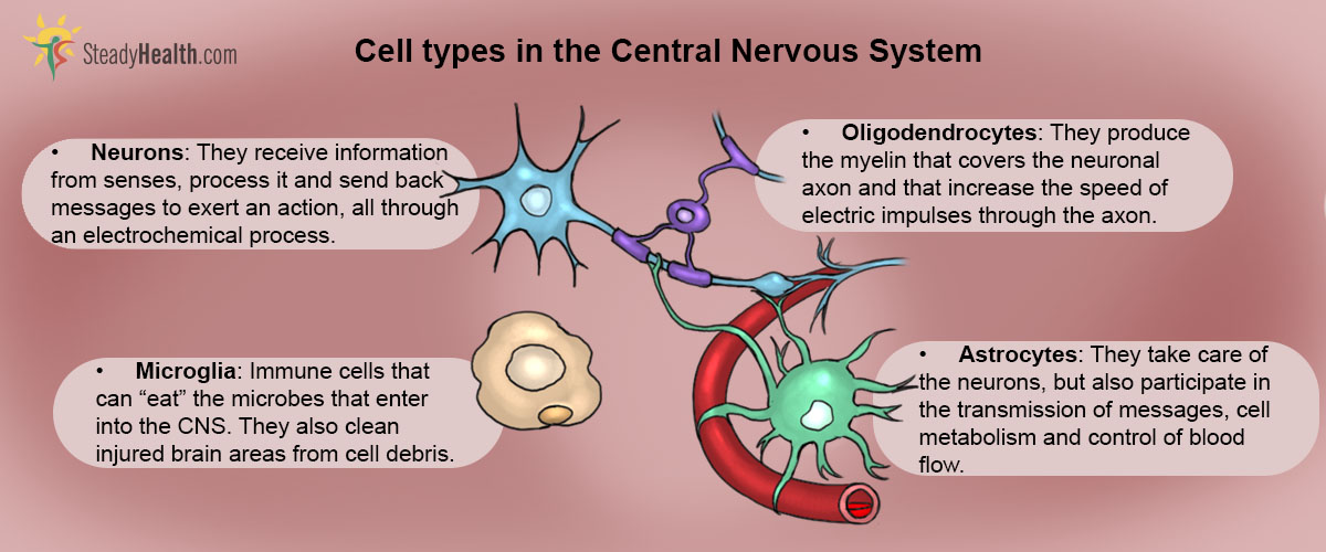
The Central Nervous System (CNS)
The CNS comprises the brain and the spinal cord. An average adult human brain weights between 1.3 to 1.4 kg and is divided in two parts, or hemispheres: the left and the right hemispheres. This division is used mainly to simplify the study of the brain, because both hemispheres are connected through a group of nerve fibres located right in the middle, known as corpus callosum.
Electric cells: Neurons
Neurons are the building blocks of the CNS. It was thought that the number of neurons in the human brain was around 100 billion, but a recent study performed in 2009 in the Universidade Federal do Rio de Janeiro, scientists estimated that the number of neurons was close to 86 billion, which is a similar to the number of neurons present in the brain of other non-human primates, like chimps.
The top of the tree is composed by the cell body from which one or more ramifications project; these projections are known as dendrites. The trunk of the tree would then be the axon, a thicker and larger structure that comes out from the cell body and ends up in what is known as the axon terminal, and looks a lot like the roots of a tree.
Nerve terminals located in every corner of our body, including our internal organs, gather the information that our body collects through our senses.
See Also: Science Maps The Connections And Functions Of Nerve Cells In The Brain Together For The First Time
It is known that an electric impulse travels at a speed of 0.1 to 100 m/s, which allows a very fast response to any stimulus.
Neurons gather all the information, process it and send a response back to where it is needed. For example, when you touch something very hot, the temperature stimulus activates receptors on your skin; these receptors send a message to the brain, which analyses the information and tells your hand muscles to move and get away from whatever is burning you.
Neuronal Carers: Glial Cells
For several years, it was thought that neurons were the most important cells present in the CNS, giving the other brain cell types, known as glial cells, the secondary role of neuronal supporters. This is still true, but scientists have discovered that non-neuronal brain cells also perform other very important functions in the CNS and that they are more than neuronal carers.
Like stars in the sky: Astrocytes
Astrocytes, as their name suggests, are star-shaped brain cells. They outnumber neurons by almost fivefold and are present all over the CNS.
Apart from this, research has shown that astrocytes also participate in the synaptic transmission and the processing of information. Brain metabolism is also one of the tasks of astrocytes; they serve as storage for energetic molecules and have a very important role in the regulation of brain glucose.
A very interesting feature of astrocytes is that their structure and function changes when the CNS is damaged. This process is known as astrocytosis, and is a common sign identified in neurodegenerative diseases.
The immune system soldiers: Microglia
Even though our CNS is protected by a very efficient fortress, formed by the skull, the vertebrae, the meninges and the cerebrospinal fluid, some microbes can have very good strategies to cross this fortress and reach the CNS. What happens, then?
Our CNS is fully ready to deal with these situations thanks to microglia.
Microglia also release substances that attract other microglial cells to the injured site in order to help with what is causing problems in the CNS. When they are not activated, microglia exert surveillance activity, which allows them to act fast and prevent the CNS from being severely damaged.
Oligodendrocytes and Schwann cells
The transmission of electric impulses from neuron to neuron works just as electricity traveling through a wire. Wires are made of materials, usually copper, that allow the flow of electrons to travel in one direction. They are also covered in a non-conductive material, like plastic, that keeps the flow of electrons isolated from other conductive materials and avoids electrocutions.
Well, neuronal axons are also covered in a non-conductive material known as myelin, which keeps the electric transmission traveling through the axon at a constant speed and prevents it from being released before reaching the axonal terminal.
See Also: Right And Left Brain Personality Theory Debunked
These cells literally fold around the axons of the neurons and segregate the substance to finally create myelin sheaths. Schwann cells are the cells in charge of the myelination of axons in the peripheral nervous system (PNS).
- AZEVEDO, F. A., CARVALHO, L. R., GRINBERG, L. T., FARFEL, J. M., FERRETTI, R. E., LEITE, R. E., JACOB FILHO, W., LENT, R. & HERCULANO-HOUZEL, S. 2009. Equal numbers of neuronal and nonneuronal cells make the human brain an isometrically scaled-up primate brain. J Comp Neurol, 513, 532-41
- GRAÇA, D. L., BONDAN, E. F., PEREIRA, L. A. V. D., FERNANDES, C. G. & MAIORKA, P. C. 2001. Behaviour of oligodendrocytes and Schwann cells in an experimental model of toxic demyelination of the central nervous system. Arquivos de Neuro-Psiquiatria, 59, 358-361
- KETTENMANN, H., HANISCH, U. K., NODA, M. & VERKHRATSKY, A. 2011. Physiology of microglia. Physiol Rev, 91, 461-553
- SOFRONIEW, M. V. & VINTERS, H. V. 2010. Astrocytes: biology and pathology. Acta Neuropathol, 119, 7-35.
- Mindmap by steadyhealth.com
- Photo courtesy of mark Miller by Flickr : www.flickr.com/photos/neurollero/51970885
Your thoughts on this