Since my father had some problems I had to be involved in some medical themes I wouldn’t be interested otherwise. He was complaining about low back pain. His doctor explained me it is necessary to do discogram. It will determine which disc has structural damage and whether it is causing his pain. I understand a lot of his diagnose, but I would like to know more about discogram, how is it done and possible risks.
Loading...
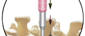
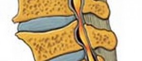
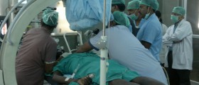
Discogram is an enhanced X-ray examination of inter vertebral discs. Dye is injected into the center of the suspected or injured disc. It makes the disc clearly visible on X-ray film and on a fluoroscope. This test is used to show if a disc has begun to rupture and if it has tears in tough outer ring. Injected fluid increases pressure, so the doctor can tell if it is painful. Your father will be given medication to help him relax. Then a long needle will be inserted from the back, which goes into the nucleus pulposus. Doctor can watch the screen as the needle is inserted to make sure it goes into the correct disc space. The risks which are associated with a discogram include infection inside the disc or allergic reaction. Also, discogram require X-rays, and those in large doses can increase the risk of cancer. Only patients who must have large number of X-rays should be concerned about this.
Loading...


