
Idiopathic Intracranial Hypertension is one specific condition characterized by high pressure in the fluid around the brain. It is also known as pseudotumor cerebri because there are some of the signs and symptoms of a brain tumor without a brain tumor being present at all. Pseudotumor cerebri literally means false brain tumor because it's symptoms mimic those of brain tumors.
What happens exactly? The fact is that the space around the brain is filled with water-like fluid called liquor! Normally- if there is too much of this fluid present, the pressure around the brain rises. This can't be avoided because the space containing the fluid cannot expand.
The condition occurs more frequently in women than men, particularly in pre-menopausal obese women. It is rare in infants.
Incidence of the condition
The fact is that BIH is a very rare condition, and is also known as Pseudotumor Cerebri (PTC) or Idiopathic Intracranial Hypertension. It affects about 2 people in every 100,000, mostly women, but can also occur in men and children. Studies of American-based populations have estimated that the incidence of pseudotumor cerebri ranges from 0.9-1.0 per 100,000 in the general population.
Possible causes of benign intracranial hypertension
Many conditions have been linked to high intracranial pressure, in fact any disorder that blocks the flow of spinal fluid between the brain and its route to the blood, the jugular vein, can cause raised pressure. The fact is that no one really knows what is causing this strange case of cranial hypertension. This is unclear, but can be often associated with:
- menstrual problems,
- hormonal problems,
- being overweight,
- certain medications- oral contraceptive and vitamin A, as well as steroid withdrawal
Symptoms of pseudotumor cerebri
It is this high pressure that produces the symptoms of idiopathic intracranial hypertension. Some of the most common symptoms are:
Headache is present in nearly all patients with Idiopathic intracranial hypertension and it is usually severe and daily.
They are different from previous headaches in life because they may awaken the patient and usually last hours, it is commonly accompanied with nausea and patients often describe it as the worst head pain ever experienced.
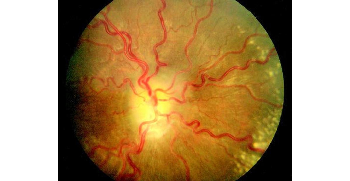
- Transient visual obscurations or blurring (68%)
What exactly are these visual obscurations? These are episodes of transient blurred vision that usually last less than 30 seconds and are followed by full recovery of vision. Visual obscurations occur in about 3/4 of IIH patients. The attacks may be involve one or both eyes. They are not correlated with the degree of intracranial hypertension or with the extent of optic nerve swelling. Visual obscurations do not appear to be associated with poor visual outcome.
- Pulse synchronous tinnitus in the ear (58%)
Pulse intracranial noises or pulse-synchronous tinnitus is very common symptoms of intracranial hypertension. In patients with intracranial hypertension, compression of the jugular vein on the side of sound abolishes it.
Patients who present with double vision most frequently complain of horizontal displacement of the images.
- Pain behind the eye (44%)
- Visual loss (30%)
- Pain with eye movement (22%)
- Shoulder and/or neck pain
- Vomiting
- memory problems
- Migraine attacks with unexplained triggers
Risk factors for developing intracranial hypertension
Exogenous substances associated with idiopathic intracranial hypertension include:
- Non-steroidal anti-inflammatory drugs (NSAIDs), tetracycline, nitrofurantoin, Isotretinoin, Tamoxifen, nalidixic acid, lithium, and the starting or stopping of steroids
- amiodarone,
- antibiotics,
- carbidopa,
- Levodopa,
- chlordecone,
- corticosteroids,
- cyclosporine,
- danazol,
- growth hormone,
- Indomethacin…
The following diseases have been associated with idiopathic intracranial hypertension:
- anemia,
- chronic respiratory insufficiency,
- familial Mediterranean fever,
- hypertension,
- multiple sclerosis,
- polyangiitis overlap syndrome,
- psittacosis,
- Reye syndrome,
- sarcoidosis,
- Systemic lupus erythematosus…
- Disorders of cerebral venous drainage
It is proven that venous compression by extra-vascular tumors or secondary thrombosis results in impaired absorption of the cerebrospinal fluid leading to pseudotumor cerebri.
Several researches done in the past have proven that pregnancy is occasionally associated with idiopathic intracranial hypertension.
Secondary Intracranial Hypertension
While pseudotumor cerebri or primary intracranial hypertension are idiopathic- secondary Intracranial Hypertension always has a cause. Possible causes include:
- dural venous sinus thrombosis
- kidney failure
- Leukemia
- Lupus
- excess Vitamin A
- growth hormone treatments
- nasal fluticasone propionate
Diagnosis of pseudotumor cerebri
The diagnosis of intracranial hypertension is made by identifying the typical symptoms of the disease along with documentation of a high spinal fluid pressure.
Recommended blood tests are:
- Complete blood count
- Erythrocyte sedimentation rate
- Serum iron and iron binding capacity
- Anti-cardiolipin antibodies/lupus anticoagulant
- Antinuclear antigen (ANA) profile
Cerebrospinal fluid studies
- Opening pressure
- White blood cell and differential counts
- Red blood cell count
- Total protein
- Quantitative protein electrophoresis
- Glucose
- Aerobic bacterial culture and sensitivity
A CT scan, or MRI on the head and brain should be done although the result is usually normal. Sometimes the ventricles may appear smaller. The neurological examination is normal except for the presence of swollen optic nerves called papilledema.
Treatment of pseudotumor cerebri
Treatment for patient with intracranial hypertension can be divided into:
- medical treatment
- surgical treatment
Medical treatment
The cornerstone of medical treatment is weight loss. It does not appear to be the total number of pounds lost. Some patients are effectively treated by losing one pound every week or two for several months and then maintaining the weight loss. It may be the loss of fluid accompanying weight loss that is the significant factor but this has not been proven.
Loss of fluid can also be obtained using diuretics (fluid pills).
Diamox (acetazolamide) is the most commonly used medication. It is relatively safe but nearly all patients have tingling of the fingers and toes. Patients also experience that carbonated soft drinks taste metallic. Another diuretic commonly used that appears to be effective in some patients is Lasix (furosemide).
Corticosteroids are also very effective in lowering the intracranial pressure in those patients with idiopathic intracranial hypertension.
Surgical treatment
- Optic nerve sheath fenestration
It is about making slits in the optic nerve sheath or covering. This procedure is only used when patients do not respond adequately to medical therapy. The operation of fenestration is done first by an incision into the orbit. The eyeball is moved to the side and the optic nerve sheath is exposed. Slits or a large hole are then placed in the optic nerve sheath and fluid drains out, thereby taking pressure off the optic nerve. Complications related to this procedure include:
- diplopia,
- optic nerve injury,
- vascular occlusion,
- a tonic pupil,
- the inherent risk of hemorrhage
- infection
Cerebrospinal fluid diversion procedures
Two neurosurgical interventions are highly effective in lowering the intracranial pressure: lumbo-peritoneal and ventriculo-peritoneal shunt! Lumboperitoneal shunts drain from the lumbar spine to the peritoneal cavity, while ventriculoatrial shunts run from the cerebral ventricles to the heart.
They represent the procedures of choice for treating patients with idiopathic intracranial hypertension who do not respond to maximum medical treatment. Unfortunately, these procedures have some complications and the most severe one being some patients have periodic occlusion of the tubing with recurrence of symptoms and sometimes vision loss.
Appropriate diet
There is no doubt that weight reduction is definitely an important factor in the long-term management of these patients. Although not too many people know more about it- as little as a 6% decrease in the total body weight can result in the resolution of papilledema. Unfortunately, weight loss in patients who are obese is difficult.
Preventing visual loss
Several researches have proven that the best way to prevent visual loss is to test vision regularly. Patients should be followed frequently with tests of vision until the doctor is confident that there is no vision loss occurring. What's bed about all this –intracranial hypertension is a life-long disease and tends to occur during periods of weight gain. The symptoms though are very treatable and, if treatment is started early enough, the vision loss is reversible.
Prognosis
Good thing about it is that prognosis is good for most patients, after treatment. The fact is also that a few will suffer with the condition becoming chronic. In this case, long term medication may be sufficient to control the condition.