Overview
The area of involvement would be similar to the area of fish that develop into gills so are located on the lateral sides of the neck if they develop.
The first branchial cleft develops into the external auditory canal but the other three remaining arches are supposed to obliterate and form no persistent structures after normal development has taken place.
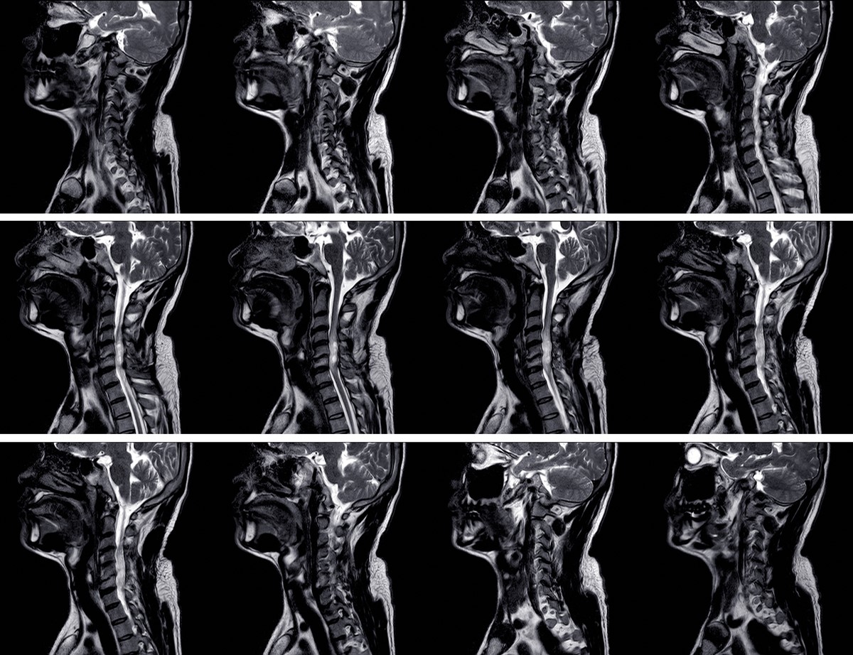
It is very important to be able to correctly diagnose any persistent and enlarging masses in children as other possible causes such as lymphoma need to be ruled out. Another issue is that these cysts can become infected and cause increased morbidity for the patient.
It is therefore imperative that these children are managed appropriately to reduce any complications that can be caused by a branchial cleft cyst.
Causes
The usual cause of a branchial cyst is the failure of obliteration of the second branchial cleft during fetal development. In other words, there is a failure of fusion of the second and third branchial arches.
Other branchial cysts may also develop due to issues involving the first, third or fourth clefts meaning that the position of the cysts will also differ.
Types
There are different types of cysts that can form due to the respective positions of each cleft.
- First branchial cleft cysts which are rare.
- Second branchial cleft cysts which, as mentioned, are the most common. They are located along the anterior border of the sternocleidomastoid muscle.
- Third and fourth branchial cleft cysts are also rare.
Symptoms
Most branchial cleft cysts are reported as slowly enlarging neck masses that are smooth. They tend to enlarge after the affected individual has contracted an upper respiratory tract infection.
The fistulas, if they are present, usually don't cause any issues but they may become infected.
Treatment
Conservative
Patients and their doctors can wait and see if the cyst ever does enlarge or cause any problems. If they do cause pain or become a cosmetic issue for the patient, then surgical intervention will be considered.
Surgery
Surgical excision will be the therapy of choice if the mass causes symptoms.
If the cysts ever end up passing near important structures such as the facial nerve, internal jugular vein, or the carotid artery then this makes complete removal of the mass a difficult situation. In such cases, as much of the mass will be removed without injuring the mentioned tissues and the patient will be followed up accordingly to monitor any changes.
It is essential then that surgeons performing these procedures understand the embryology and anatomy of these masses so that they can provide adequate treatment to affected patients.
- www.sempedsurg.org/article/S1055-8586(06)00014-X/fulltext
- Photo courtesy of SteadyHealth