
Teeth (Chewing)
- Important notification about information and brand names used in this slideshow!
- Photo courtesy of Stacey Huggins by Flickr : www.flickr.com/photos/staceyhuggins/5055677838/
- Cunningham's manual of practical anatomy

Salivary Glands
- Important notification about information and brand names used in this slideshow!
- Photo courtesy of allnightavenue by Flickr : www.flickr.com/photos/allnightavenue/6469511099/
- Cunningham's manual of practical anatomy

Esophagus
- Important notification about information and brand names used in this slideshow!
- Photo courtesy of Dan Zen by Flickr : www.flickr.com/photos/danzen/981445584/
- Cunningham's manual of practical anatomy
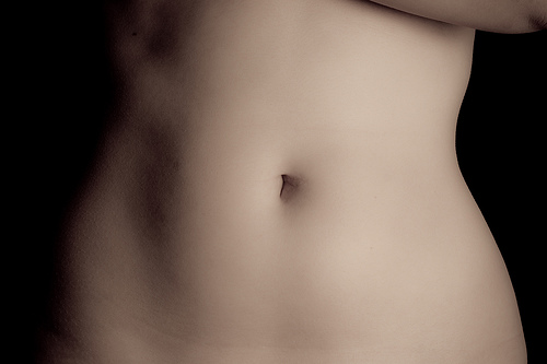
Stomach
- Important notification about information and brand names used in this slideshow!
- Photo courtesy of Filip Bunkens by Flickr : www.flickr.com/photos/loneblackrider/3175090147/
- Cunningham's manual of practical anatomy
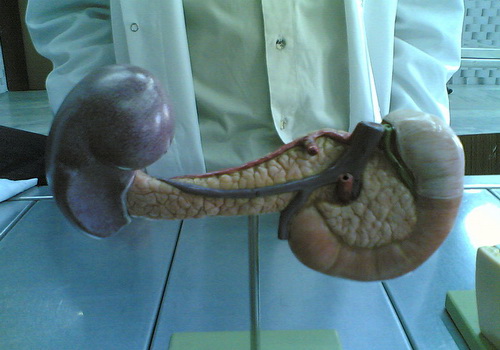
Pancreas
- Important notification about information and brand names used in this slideshow!
- Photo courtesy of Süleyman Habib by Wikimedia Commons : commons.wikimedia.org/wiki/File:Pancreas_model_back.jpg
- Cunningham's manual of practical anatomy
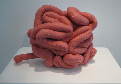
Large Intestines
- Important notification about information and brand names used in this slideshow!
- Photo courtesy of Hey Paul Studios by Flickr : www.flickr.com/photos/hey__paul/8480135160/
- Cunningham's manual of practical anatomy
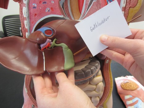
Gallbladder
- Important notification about information and brand names used in this slideshow!
- Photo courtesy of ursulaar by Photobucket : media.photobucket.com/user/ursulaar/media/IMG_0597.jpg.html?filters[term]=gallbladder&filters[primary]=images&sort=1&o=34
- Cunningham's manual of practical anatomy
Liver
- Important notification about information and brand names used in this slideshow!
- Photo courtesy of Darren Barefoot by Flickr : www.flickr.com/photos/dbarefoot/17083596/
- Cunningham's manual of practical anatomy
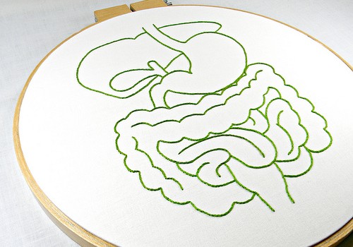
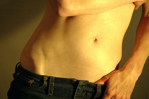
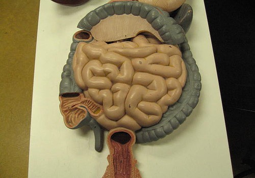
Small Intestines
- Important notification about information and brand names used in this slideshow!
- Photo courtesy of Cliff by Flickr : www.flickr.com/photos/nostri-imago/2849341081/
- Cunningham's manual of practical anatomy
Rectum And Anal Canal
- Important notification about information and brand names used in this slideshow!
- Photo courtesy of GreenFlames09 by Flickr : www.flickr.com/photos/greenflames09/143141020/
- Cunningham's manual of practical anatomy
Your thoughts on this
Loading...