RIGHT TESTIS measuring 3.3 x2.0cm. *normal in size, shape withsmooth outer surface. *The echogenecity of the parenchyma seen normal. *No evdence of focal mass seen. *Epididymus seen normal. *No cystis tubular structures seen adjacent to the testis.
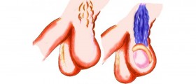
LEFT TESTIS measuring 3.7x1.9cm *normal in size, shape withsmooth outer surface. *The echogenecity of the parenchyma seen normal. *No evdence of focal mass seen. *Epididymus seen normal. *tiny cyst formed in the head of left epididmis measure 0.1 x 0.1cm (TSXAP) cyst having thin walled showing clear fluid. *tortuousily of peri-testicular vessels lateral and posterior to the left testis, maximum diameter upto 2.5mm represents varicocelle. However no definite evidence of retrograde flow seen on valsalve meneuver.
CONCLUSION: tiny cyst formed in the head of left epididmis measure 0.1 x 0.1cm (TSXAP) cyst having thin walled showing clear fluid representing spermatocelle and tortuousily of peri-testicular vessels lateral and posterior to the left testis, maximum diameter upto 2.5mm represents varicocelle. However no definite evidence of retrograde flow seen on valsalve meneuver. No evdence of mass and hydrocelle seen in either both side. Both testes apparently normal. Clinical correlation would be suggested.
MY DOCTOR SAID TO ME; IT IS NOT DANGEROUS IT'S JUST SPERMATOCELE. IS HE RIGHT?
Loading...


