Anatomy of Brainstem


In general, the brainstem deals with communications between the rest of the brain and the body and the more primitive (but still essential) functions such as the vasomotor, respiratory and cardiac systems.[1]
The brain stem is the stalk of the brain below the cerebral hemispheres. It is made up of three different parts or segments[1]:
- The upper segment of the human brain stem is called the pons. It contains nerve fibers that connect the two parts of the cerebellum. That’s why it is vital in coordinating movements of the body.
- The second part of the brain stem is positioned below the pons and continuous with the spinal cord. This part is called the medulla. The main function of this part is a transmission of ascending and descending nerve fibers between the spinal cord and the brain.
- The nuclei of some of the nerves that originate in the brain are also located in the brain stem.
- Sometimes the diencephalon, the caudal part of the forebrain, is included, too.
Function of brainstem
The brainstem controls many functions. Besides the fact that it represents a major route for communication between the forebrain, the spinal cord, and peripheral nerves, it also controls various autonomic functions such as respiration and heart rhythm as well as perceptual functions such as the primary aspects of sound localization. Neurological functions located in the brainstem include those necessary for survival such as breathing, digestion, heart rate, blood pressure and for arousal such as being awake and alert. It also contains most of the cranial nerves.
Parts of the brainstem
- Medulla Oblongata
Medulla oblongata functions primarily as a relay station for the crossing of motor tracts between the spinal cord and the brain. It also contains the respiratory, vasomotor and cardiac centers, as well as many mechanisms for controlling reflex activities such as coughing, gagging, swallowing and vomiting. Therefore medulla oblongata represents one of the most important parts of the brainstem.
- Midbrain
The midbrain is the highest part of the brainstem and it serves as the nerve pathway of the cerebral hemispheres. It is proven that it contains auditory and visual reflex centers.
- Pons
The pons is the middle part of the brainstem and it represents a bridge-like structure which links different parts of the brain and serves as a relay station from the medulla to the higher cortical structures of the brain. The most important center that the pons contains is the respiratory center.
- Long tracts which are passing through the brainstem
Long tracts, the motor and sensory tracts described in the spinal cord are present in the brain stem, but in the brain stem, they are all contralateral to the side of the body they serve.
- The pyramidal tract
The pyramidal tract is located in the cerebral peduncles of the midbrain. It courses through the base of the pons in several bundles that rejoin to form the pyramids in the medulla. The pyramids decussate at the junction of the medulla and cervical spinal cord.
- The spinothalamic tracts
The spinothalamic tracts continue in a lateral position throughout the brainstem on their way to the thalamus. The axons from the 1st order neuron located in the dorsal root ganglion enter the dorsal root entry zone and within several segments synapse with 2nd order neurons in the dorsal horn. Axons from the 2nd order neuron ascend as the spinothalamic tract to the ventral posterior lateral nucleus (VPL) of the thalamus. The axons of the 3rd order neurons project to the Somatosensory cortex.
- Dorsal Column-Medial Lemniscus
The axons from the 1st order neurons located in the dorsal root ganglion ascend in the dorsal columns on the same side of the cord until they reach the 2nd order neurons in the medulla. Axons from the 2nd order neurons cross at the level of the medulla and then travel near the midline in the medial lemniscus. The 3rd order neuron projects to the primary Somatosensory cortex.
Types of brainstem lesions
All brainstem lesions could be divided into two big groups [2, 3] :
1. Primary Brainstem Lesions
Primary Traumatic Effects
NEURAL
- Hypoxic neurons (focal)
- Neuronophagic cell death
- Axonal injury (focal / diffuse)
VASCULAR
- Focal edema
- Small hemorrhages
- Hematoma
- Damage to large vessels
- Necrosis
DELAYED EVENTS
- Cell dysfunction
- Differentiation
- Chromatolysis
- Trans-neuronal degeneration
- Late cell death, apoptosis
- Tract-degeneration
- Re-Innervations after axonal injury
2. Secondary Traumatic Effects
- Swelling or edema
- Global ischemia
- Increased ICP or herniation of cerebral tissue
- Haematoma and hemorrhages of different size
Classic syndromes related to the brainstem lesions
There are several syndromes that appear after lesions in brainstem region of the brain and they are called the brainstem syndromes.
Mid-brain syndromes
- Weber's syndrome
The critical structures involved are the two most important pathways - descending corticospinal and corticobulbar fibers in the cerebral peduncle, and the fibers of the third nerve that traverse the peduncle on exiting the midbrain. If the lesion is on the right; the patient will have a left hemiparesis and a right third nerve palsy and vice versa.
- Benedikt's syndrome
Midbrain stroke involving the third nerve is causing this syndrome characterized by third nerve palsy on one side with contralateral ataxia.
Medial pons syndromes
It is proven that lesions of the sixth nerve nucleus cause paralysis of gaze to the side of the lesion. Lesions involving the fibers of the sixth nerve as they travel through the pons can also involve the medial lemniscus. This is causing unilateral abducens weakness and contralateral loss of position and vibration.
Lateral medullar syndrome (Wallenberg syndrome)
This is the most common of the brain stem strokes. When this stroke involves the spinothalamic tract, it results in contralateral loss of pain and temperature sensation below the neck. Involvement of the descending nucleus and tract of V results in loss of pain and temperature sensation on the face ipsilateral to the lesion.
The locked in syndrome (infarction of the base of the pons)
In this syndrome the corticospinal and corticobulbar tracts in the basis of the pons are interrupted which leads to the quadriplegia and paralysis of all cranial nerve muscles except for those controlling eye movements. This means that the person is literally “locked in”, accept their eyes. If the lesion extends into the tegmentum of the caudal pons, horizontal eye movements may also be affected while vertical eye movements are still possible, and sensation can be affected. In this case the only way to communicate with these unfortunate patients is to ask them to move their eyes in response to questions.
- Pontine hemorrhage
Bleeding or hemorrhage into the pons results in coma, decerebrate posturing, and small pupils.
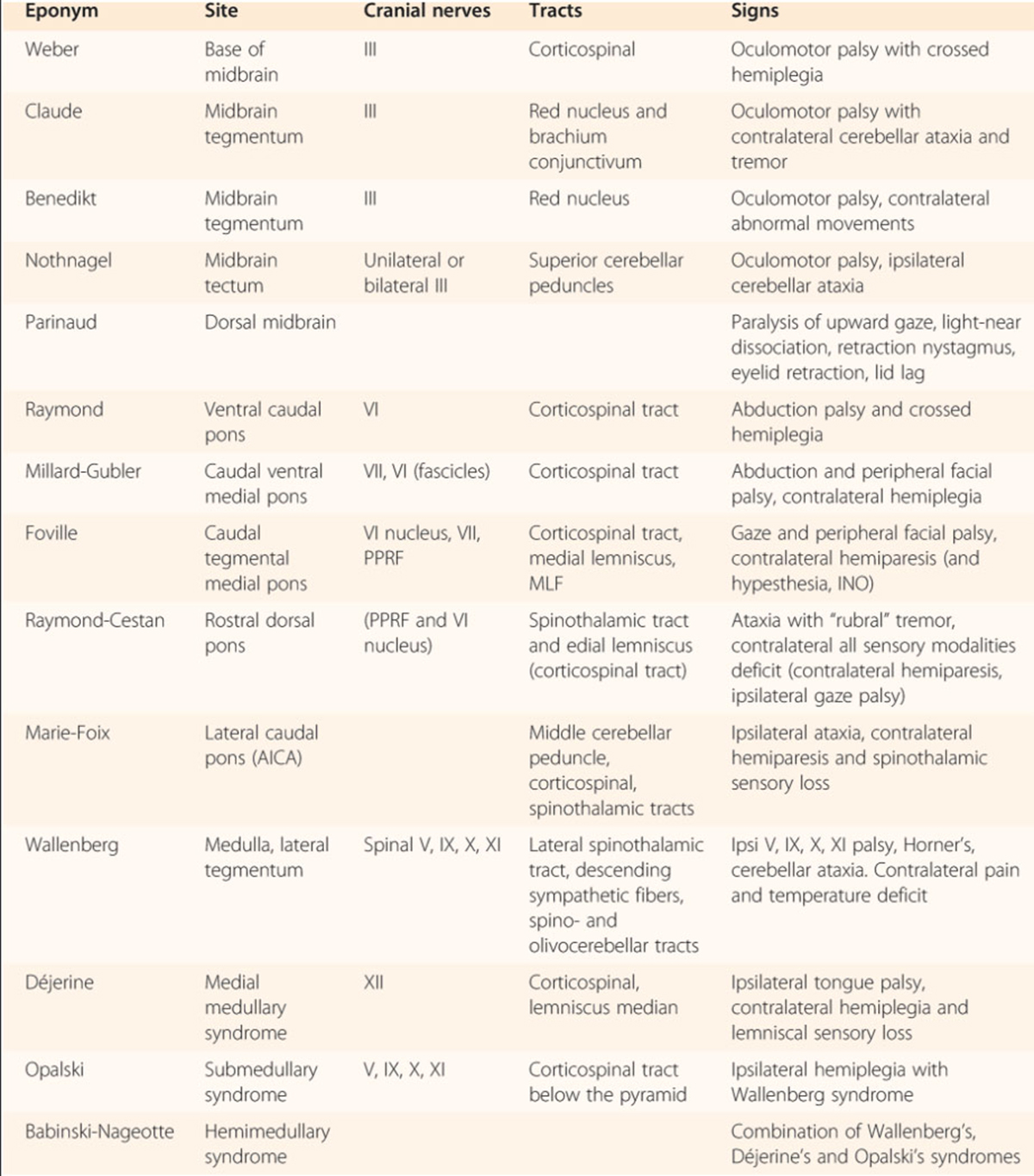
Other less common brainstem syndromes [4]
Cranial nerves and brainstem
All cranial nerves except I and II, originate in the brainstem, which includes the midbrain, the pons, and the medulla. There are 12 cranial nerves and they can be divided into sensory, motor, or mixed nerves. Overall, sensory nerve nuclei tend to be located in the lateral brainstem, while motor nuclei tend to be located medially.