Limited blood flow usually ensues as a result of atherosclerosis, a process where plaque (made of calcium, fat, and cholesterol) slowly builds up on your arterial walls. Plaque narrows the lumen of the artery, which restricts the flow of blood circulating through your vessels, meaning that the organs do not receive enough oxygenated blood.
The effects of this process are most frequently seen in your neck arteries and your legs. However, aortic blood flow may also be compromised via this mechanism. The abdominal part of the aorta, specifically the part below the renal arteries, is particularly affected. This is called aortic occlusive disease. If the iliac arteries are also affected by this process, it is known as aortoiliac occlusive disease.
How do I get aortic occlusive disease?
Other than atherosclerosis, aortic occlusive disease can also be caused by some forms of inflammatory diseases of the aorta, such as Takayasu arteritis, giant cell arteritis, or arteritis caused by ionizing radiation.
Other causes of aortic occlusive diseases include:
- Fibromuscular dysplasia, where the arterial wall grows so thick that it narrows the lumen.
- Aortic dissection, characterized by a tear in the inner layer of the aorta.
- Coarctation of the aorta, a birth defect where the aorta is narrower in some areas.
- Aortic atresia, also a birth defect in which the aortic valve on the heart is missing.
Risk factors for aortic occlusive disease are mostly the same as in all arterial diseases:
- Smoking, as almost two thirds of patients are male smokers.
- High levels of fat.
- Obesity.
- High blood pressure.
- Diabetes.
- Low levels of physical activity.
Aortic occlusive disease progresses very slowly. During that process, blood may bypass its path via collateral circulation, which includes a network of small blood vessels which are not normally open. However, since the aorta is the largest artery in your body, no amount of collateral circulation can entirely make up for the blockage of the aortic pathway. In these cases, the extent of aortic insufficiency entirely depends on how much the collateral system in that area is developed.
What are the signs and symptoms of aortic occlusive disease?
In most cases, aortic occlusive disease is characterized by a group of symptoms called Leriche syndrome, which involves:
- Absence of pulse in the groin region.
- Pain or cramping in the lower part of the legs due to decreased blood flow, called claudication. It can appear either during physical activity, or if the condition is serious, even during rest.
- Erectile dysfunction (impotence).
It’s most commonly diagnosed in older patients who already have other chronic diseases, such as malignancies, arrhythmia, lung, kidney, or neurological diseases. Because of decreased blood flow, blood clot formation on the plaque is common. The blood clot may eventually break off and cause an acute clog (manifesting in the so-called blue toe syndrome, or in severe cases, even cut off the blood flow to the entire lower half of the body.
How is aortic occlusive disease diagnosed?
During a physical exam, your doctor may check the pulse on your ankle. However, measuring blood pressure in segments is a more precise approach. You might be asked to walk on a treadmill, so your doctor can evaluate under what levels of physical activity claudication starts, and after how much time.
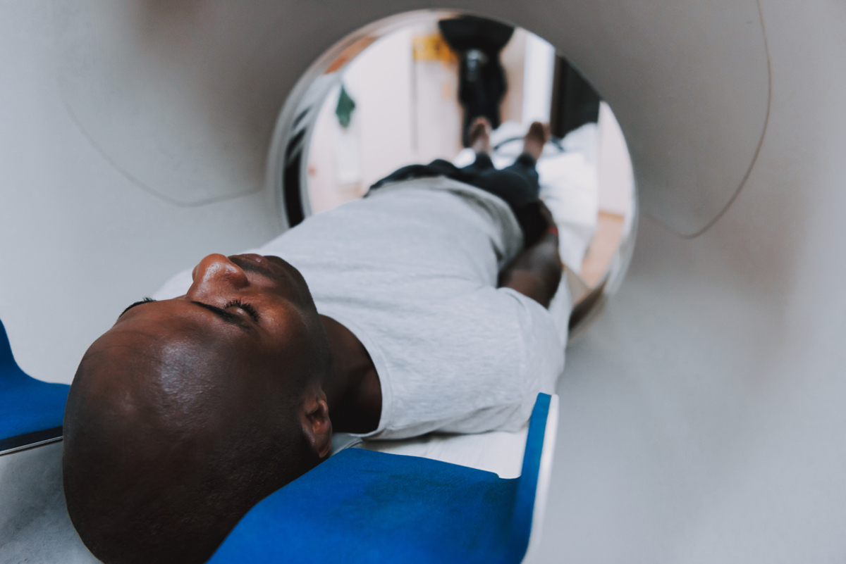
Imaging plays a crucial role in precisely diagnosing occlusions. Imaging procedures involve:
- Ultrasound, or doppler ultrasonography, to be more precise. Besides traditional ultrasound, on which you can view and measure the dimensions of organs and blood vessels, doppler sonography detects and shows the blood flow and its properties. Nowadays, modern ultrasound machine can show regular ultrasound pictures along with blood flow visualization, and that method is called duplex sonography.
- CT scan, which can visualize the aorta in great detail, detecting plaques and aneurysms, with the potential to discover other abdominal or pelvic irregularities
- MRI angiography, which gives similar information as a CT, although it’s more suitable for patients with kidney issues, because of the difference in contrast agents. Also, unlike CT, MRI doesn’t include ionizing radiation. However, the method is still very expensive.
- Aortography, where a contrast agent is injected via a catheter placed in the aorta. It gives much information about the blood vessels, although it’s not suitable for all patients, and is recommended only when surgery is necessary.
It is also important to differentiate claudication caused by aortic occlusive disease and the one caused by the narrow spinal canal. This can be done during an MRI scan.
How to treat aortic occlusive disease?
If you have claudication during rest, or initial necrotic changes, which if untreated can lead to amputation of the limbs, surgery is in order.
The most effective surgical procedure is the aortobifemoral bypass, where the prosthetic “Y” shaped replacement is positioned in place of the problematic part of the aorta and the iliac arteries.
If, for some reason, this procedure is impossible, doctors can do an axillobifemoral bypass, done in local anesthesia. It involves inserting a plastic tube under the skin, which connects the axillary artery in your shoulder with the femoral arteries in your legs.
A “cross-over”, or femorofemoral bypass is indicated if one of the iliac arteries is normal. The graft connects the femoral artery from the healthy with the same artery on the other, receiving side. Aortic thromboendarterectomy (TEA) is a procedure rarely used today, which includes removal of the blood clot from the arterial vessels.
Endovascular surgery is growing in presence nowadays. This minimally invasive procedure involves placing of a synthetic graft in the problematic area. It is used to treat stenosis no longer than 5 centimeters in length.
The goal in aortic occlusive disease treatment is to slow down the progression of the disease, while simultaneously stimulating collateral circulation to develop, and preventing muscle atrophy. This can be done using medications such as Aspirin or Clopidogrel, which reduce the risks of complications.