Dental implants have become common enough that many patients know about them even before walking into a dental clinic. The dentist cannot (as much as they would like to), however, place a dental implant in every patient that walks through the door!
What is a CT scan?
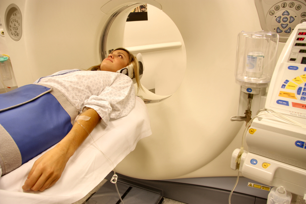
Computed tomography scans, also known as CT scans, are special types of x-rays. Taken with the help of a CT scanner, this imaging technique takes a series of x-rays of the body, from a variety of different angles. Then, using the processing power of a computer, these x-rays are stitched together to form a cross-sectional image of the organs inside the body.
These cross-sections are known as "slices". They give a more detailed view of the bones, blood vessels, nerves, and other soft tissues inside the body, compared to conventional x-rays.
How are CT scans used in implant dentistry?
Treatment planning is the key to a successful implant procedure. There are many tools that can help a dentist plan implant surgery. Previously, intraoral x-rays and OPG’s were used to determine the amount of bone present in a patient’s jaw. But these provide a two-dimensional view of a three-dimensional structure — the jaw bone.
That's where CBCT comes in
Cone Beam Computed Tomography (CBCT) is now extensively used in dentistry, particularly implant dentistry. It is a special type of CT scanner that uses conical beams to generate a three-dimensional image of the jaw bone. This image helps the surgeon to accurately visualize the amount of bone present in the jaw, the numerous critical nerves and vessels passing through it, as well as its density.
How is a CBCT done?
The patient is typically asked to sit upright on a chair while the technician centers the machine over the area of interest. The patient is required to sit still, while the machines rotate 360 degrees around the patient’s head, capturing images from different angles, and giving a three-dimensional picture. The entire process typically lasts less than a minute.
CBCT machines have reduced in cost and some bigger dental practices have started keeping a dedicated CBCT machine as a part of their setup.
Precautions you should take before a CBCT
In order to get a clearer and more accurate image, you will be asked to take off any accessories like earrings, necklaces, spectacles, etc. prior to the scan. If a patient is wearing any removable prosthesis like dentures, they need to be taken off too.
Advantages of getting a CBCT before implant surgery
Implant size selection
A CBCT can give the exact dimensions of the bone, helping the surgeon select the ideal diameter and length of the implant, as well as select the site for implant placement.
Avoid complications
A CBCT gives the exact position of the blood vessels, sinuses, and major nerves present in the jaw, helping the surgeon to bypass these vital structures during the surgery and avoid complications. It can also help the surgeon to determine the angulation of the implant in such a way that the implant stays centered within the jaw bone.
Determining the need for additional surgeries
In case the bone dimensions are found to be insufficient, the surgeon can plan to carry out additional surgeries like sinus lifts and ridge splits during or before the implant surgery.
Is getting a CBCT risky?
While a CBCT does expose you to more radiation than a conventional intraoral x-ray, this radiation is highly focussed on the area being scanned, reducing the amount of scattered radiation. Getting a CBCT is safe, with almost no risks.
Surgical stents and guided surgery
Individual custom-made guides can be made using the three-dimensional image obtained by a CBCT. This guide can be easily attached to a patient’s jaw during the surgery. This guide contains holes through which the surgeon can accurately place the implant in the bone at the selected site at a predetermined depth and accurate angulation.
Is a CBCT expensive?
Dental clinics performing a large number of implant surgeries have in-house CBCT facilities and the cost of a CBCT is usually factored into the total cost of the implant procedure.
Certain radiology clinics too offer the services of a CBCT. While the cost of a CBCT is higher than that of a conventional x-ray, it has reduced considerably over time. The benefits that a CBCT provides in the form of treatment planning far outweigh the costs of getting one.
Conclusion
Successful implant surgery and a long-lasting prosthesis can only be provided if the patient’s jaw bone is dense enough and has the required height and width to support the implant.
Your thoughts on this