
What is dilated cardiomyopathy?
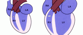
Dilated cardiomyopathy is a condition affecting the cardiac musculature. As the muscles are weakened, the chambers of the heart become dilated. The main pumping chamber of the heart, that is the left ventricle, becomes enlarged and is no longer able to push the blood into the aorta with as much force as it would have otherwise. The right ventricle may also become dilated and dysfunctional. Dilated cardiomyopathy is a progressive disease which may ultimately lead to heart failure. In fact, it is the third most common cause of heart failure and often a reason behind the need for a heart transplant.
- Important notification about information and brand names used in this slideshow!
- Photo courtesy of ceso83 by Photobucket : media.photobucket.com/user/ceso83/media/DSCN0158.jpg.html?filters[term]=chest%20pain&filters[primary]=images&filters[secondary]=videos&sort=1&o=7
- www.webmd.com/heart-disease/guide/dilated-cardiomyopathy
- http://www.medicinenet.com/cardiomyopathy_dilated/article.htm

Symptoms of dilated cardiomyopathy
Early stage of dilated cardiomyopathy usually does not produce any symptoms. Symptoms of advanced dilated cardiomyopathy are the same as that of heart failure. They include are: • Shortness of breath, especially on exertion • Swelling over feet, increase in weight and increase in abdominal girth • High blood pressure • Increased pulse rate • Increased jugular venous pressure • Wheeze • Signs of impaired oxygenation of tissues like cyanosis and clubbing • Lightheadedness and fainting • Irregular heart rhythm • Palpitations (flutter caused by irregular heart rate) • Because of pooling of blood in the left ventricle, clots may be formed. These clots may dislodge and reach the lungs causing pulmonary embolism or they may travel up to the brain resulting in stroke. • Pain in chest • The patient may die suddenly

Swelling of the lower extremities
Patients of dilated cardiomyopathy suffer from both right and left sided heart failure. As there occurs pooling of blood in the left ventricle, not enough blood is pumped into the arteries. Kidneys perceive this reduced cardiac output as a reduction in blood volume. So they retain salt and water thereby, increasing the total blood volume. The collection of blood in the veins of the feet leads to leakage of fluid from the capillaries into the interstitial space. This results in swelling of lower extremities. Moreover, reduced cardiac output results in building up of back pressure. This also causes enlarged liver and pitting edema of lower extremities.
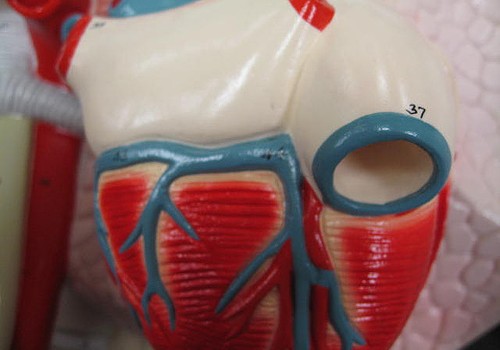
Causes of dilated cardiomyopathy
The common causes of dilated cardiomyopathy include: • Genetic: Around 20% of these cases are hereditary. • Alcoholism • Tachycardia induced dilated cardiomyopathy commonly caused by atrial fibrillation. • Drug abuse involving cocaine, heroin and methamphetamine • Prescription medicines like phenothiazines and chemotherapeutic medicines like cyclophosphamide, mitoxantrone, trastuzumab, etc. • Hemochromatosis • Coronary artery disease • HIV • Pregnancy induced, especially in women suffering from pre-eclampsia and post-partum dilated cardiomyopathy. • Infectious diseases like Rocky Mountain spotted fever, leptospirosis, Lyme disease, toxoplasmosis, Q fever, Chagas disease, cytomegalovirus, adenovirus, etc. Rarely, dilated cardiomyopathy may be caused by: • Connective tissue diseases like systemic lupus erythematosus, rheumatoid arthritis, scleroderma, polyarteritis nodosa • Endocrinal diseases like thyroid disorders, diabetes, acromegaly and obesity • Electrolyte disturbances • Toxins like cobalt, mercury and organic solvents

Severe coronary artery disease and dilated cardiomyopathy
Severe coronary artery disease is associated with building up of plaque inside the lumen of the arteries. It may also result in the hardening of the vessels. When the lumen of the arteries that bring blood and oxygen to the heart muscles (coronary arteries) gets blocked, the blood flow to the cardiac muscles becomes impaired. Over time, the functioning of cardiac muscles gets affected. So they are no longer able to contract properly. This is termed as ischemic cardiomyopathy. As the heart muscles do not contract properly, all the blood from the left ventricle cannot be pushed out and tends to pool inside. This leads to the dilatation of left ventricle, a condition called as dilated cardiomyopathy.

Alcoholism and dilated cardiomyopathy
Alcohol consumption over a long time can lead to secondary, non-ischemic cardiomyopathy. Although the exact mechanism by which alcohol causes cardiomyopathy has not been established, it has been proposed that alcohol produces the following changes: • Inhibits protein synthesis • Inhibits oxidative phosphorylation • Free radical damage • Accumulation of fatty acid esters • Disrupts the cell membrane structure of cardiac cells • Vasospasm of the coronary arteries • Receptor anomalies in cardiac cells • Activates the renin-angiotensin mechanism All these changes together lead to dilated cardiomyopathy. Apart from cardiomyopathy, chronic alcohol consumption can lead to atrial fibrillation, supraventricular arrhythmias, premature contraction of the ventricles, high blood pressure and stroke.
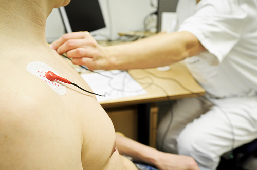
Changes in the ECG in patients with dilated cardiomyopathy
Patients suspected of having dilated cardiomyopathy are subjected to a routine electrocardiography. The ECG in such patients gives an idea about the size of the left ventricle and the other chambers of the heart. There are non-specific ST-T wave changes and Q waves seen in the ECG which are characteristic of dilated cardiomyopathy. Left bundle branch block and other conduction delays may be present. Atrioventricular blocks of different levels are also seen in the ECGs of different patients. The presence of atrial fibrillation in the ECG signifies that the disease is in an advanced stage and may be progressing to heart failure. Absence of atrial fibrillation means that there is no eminent danger of heart failure.
- Important notification about information and brand names used in this slideshow!
- Photo courtesy of Universitetssykehuset Nord-Norge (UNN) by Flickr : www.flickr.com/photos/unnfoto/6068321821/
- emedicine.medscape.com/article/152696-workup#aw2aab6b5b6
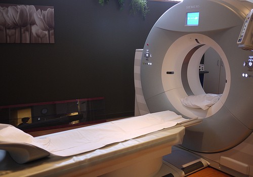
Changes seen on X-ray in dilated cardiomyopathy
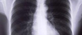
Although the diagnosis of dilated cardiomyopathy is made on the basis of several tests like electrocardiography, echocardiography, Holter study and treadmill test, the most common test which gives the physician an idea of the underlying disease is x-ray of the chest. In patients suffering from dilated cardiomyopathy, x-ray of the chest shows an enlarged cardiac shadow. The pulmonary trunk also appears to be bigger than normal. There may be evidence of pleural and pericardial effusion although this is not necessary in all the cases. In a chest x-ray, cardio-thoracic ratio (CTR) is the ratio of the transverse cardiac diameter to the maximal internal diameter of the thorax at the level of the diaphragm. A CTR more than 0.5 represents cardiomegaly, which is often seen in dilated cardiomyopathy.

Medications for dilated cardiomyopathy
Depending upon the severity of the symptoms, a patient of dilated cardiomyopathy may require a combination of two or more of the following medicines: • Angiotensin-converting enzyme (ACE) inhibitor: They dilate the blood vessels thereby lowering the blood pressure. They improve the blood flow and reduce the heart’s workload. • Angiotensin II receptor blockers: They exert the same benefits as ACE inhibitors and are devoid of the side effect of cough that the former has. • Beta blockers: They slow the heart rate and reduce the blood pressure. They help in relieving the symptoms of heart failure and enhance functioning of the heart. • Diuretics: They increase urine output. They thus, reduce the blood volume and relieve the congestion of the lungs associated with heart failure. • Aldosterone antagonists: They act as diuretics without disturbing the potassium balance of the body. • Digoxin: It slows the heart rate and increases the force of contraction. • Blood thinners: They help in preventing the formation of clots associated with pooling of blood.
Living with dilated cardiomyopathy
People with dilated cardiomyopathy have to make certain adjustments with their lifestyle. These include: • Patients shouldn’t participate in competitive sports as it increases the demand on the heart. Participating in such sports increases the risk of the heart stopping abruptly and sudden death. • Patients are recommended to give up alcohol and illegal drugs completely as they may worsen their condition. • Patients are advised to quit smoking. • Eat healthy and maintain a healthy weight. Obesity increases the pressure on the heart. A failing heart may not be able to bear with the increased demand. In order to maintain a healthy weight, patients are advised to eat a well-balanced diet and take adequate amount of rest. They can consult a dietician for this purpose. • Patients with symptoms of heart failure may have to keep in check their salt and water intake according to their physician’s advice.

















Your thoughts on this
Loading...