
What is Chondromalacia patellae?
Chondromalacia patella is a condition in which the cartilage underneath the knee cap (patella) is softened due to degenerative changes in it. These changes occur as a result of poor alignment of the patella while it slides over the distal end of the thigh bone (femur). The poor alignment leads to rubbing of the cartilage every time the patella moves over the femur. Continuous stress over the knee cap for a long duration of time, while running, jumping or playing soccer, may also lead to chondromalacia patella. It is a common cause of chronic pain in front of the knee in young people. The pain aggravates while climbing up or down the stairs.
- Important notification about information and brand names used in this slideshow!
- Photo by shutterstock.com
- www.mayoclinic.com/health/chondromalacia-patella/DS00777
- http://en.wikipedia.org/wiki/Chondromalacia_patellae
- http://www.patient.co.uk/health/chondromalacia-patellae-leaflet

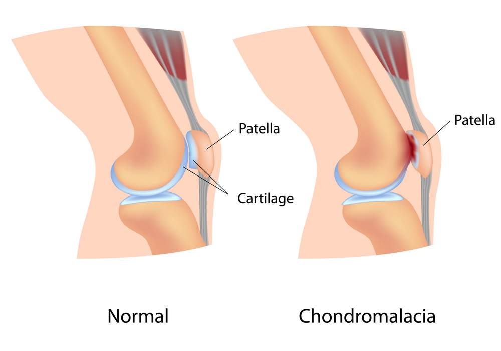
Symptoms of chondromalacia patella
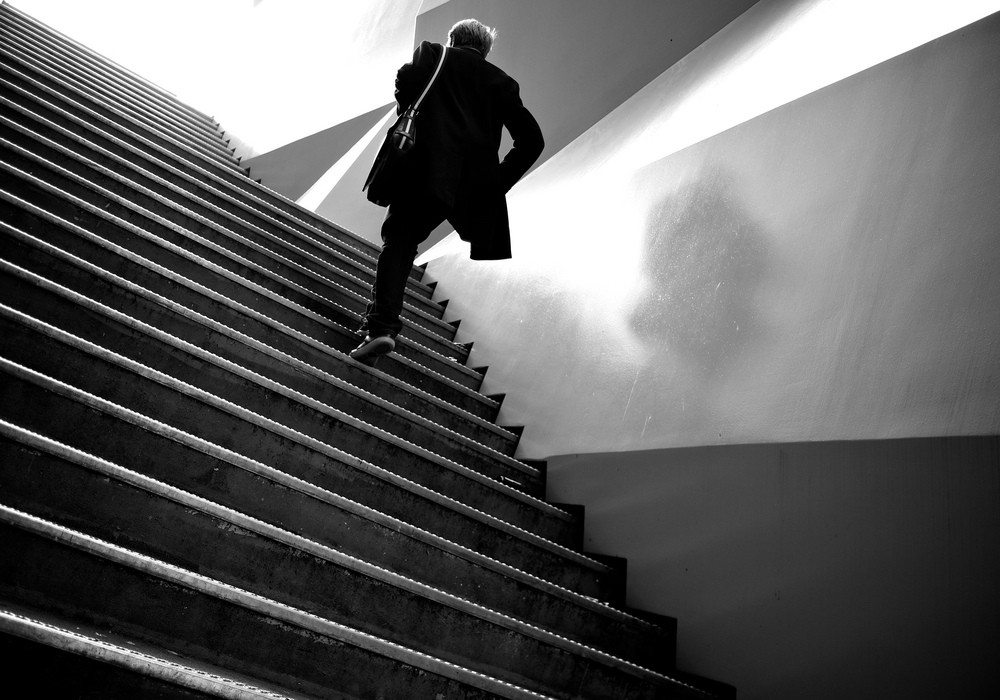
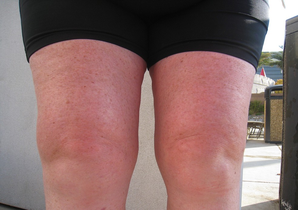
The most common symptom of chondromalacia patella is a dull aching pain on the anterior aspect of the knee joint. This pain is aggravated by ascending or descending the stairs, kneeling, squatting, or sitting with a moderately bent knee for a prolonged time (the so called theatre sign). It also worsens when pressure is put on the knee over an extended time like standing, running or heavy exercising. There is a grating or grinding sensation every time the knee is extended (crepitus). Some amount of swelling may be noted in front of the knee. In rare circumstances, there may be some effusion in the knee joint.

Causes of chondromalacia patella
In chondromalacia patella, there is a non-alignment of the tendons or ligaments working on the patella. These tendons and ligaments attach the patella to the thigh bone (femur) or the shin bone (tibia). Due to the non-alignment, every time the knee is flexed, the patella rubs against the lateral side of the femur. This constant friction between the two bones leads to damage to the articular cartilage, situated beneath the patella. It becomes inflamed and swollen. The pain arising due to the inflammation is the characteristic pain on the anterior aspect of the knee experienced by a person suffering from chondromalacia patella.

Use of knee caps
The knee cap, known as patella in medical parlance, is a movable bone situated in front of the knee. The patella is wrapped inside the quadriceps tendons which attach the quadriceps muscles to the bones of the lower leg. Tightening of the quadriceps muscles makes the tendon tense and straightens the leg. The patella acts as a fulcrum and increases the pull of the quadriceps muscles. The under-surface of the knee cap is covered by a slippery articular cartilage. This cartilage facilitates the smooth gliding movement of the knee cap over the femur. There are two muscles which help in keeping the patella in its position when the leg is straightened. If the timing between these two muscles is off, it results in the non-alignment of the patella and damage to the articular cartilage.

Who's at risk of developing chondromalacia patella?
People who routinely take part in some activity that involves constant wear and tear of the knee joint are at a high risk of developing chondromalacia patella. The condition is extremely common in young athletes. Besides athletes, chondromalacia is more likely to develop in soccer players, snow boarders and tennis players. It is also seen in ballet dancers and cyclists. Chondromalacia can develop as a part of the normal ageing process. It may also develop when there is a lot of pressure on the knee joint as in case of obesity, or in people with a flat foot. Women are twice more likely to develop the condition. This is because they have a wider pelvis which increases the angle at the meeting point of the bones in the knee joint.

Diagnosis of chondromalacia patella
Physical examination of the knee joint by an expert is the single most important step in the diagnosis of chondromalacia patella. Not only is the expert able to appreciate the degree of swelling and pain, physical examination while keeping the knee joint in various positions of extension and flexion helps him to differentiate chondromalacia form other knee conditions which may produce similar symptoms. Apart from manual examination, your physician may also like to go for: • X-ray of the knee joint: This helps him in observing the angle that the two main bones of the leg make at the knee joint. X-ray is of not much help in visualizing the soft tissue swelling. • CT scan of the knee joint: This gives a good idea of both the bones and the soft tissues. • Magnetic Resonance Imaging of the knee joint: This is the best diagnostic test to examine the knee joint but is very expensive and is often not needed in making the diagnosis of chondromalacia patella.

Medications/pain relievers for chondromalacia patella
Resting the knee joint, applying ice, medications to relieve pain and bracing the knee joint are the important initial steps to deal with chondromalacia patella. Resting the knee joint gives time for the swelling and inflammation of the joint to subside. Applying ice on the knee cap for at least 15 minutes two times a day helps in reducing the inflammation. Non steroidal anti-inflammatory medications are useful in providing relief from the severe pain and inflammation of the knee joint. Ibuprofen and Naproxen are the commonly prescribed NSAIDs in this condition. Knee brace helps in keeping the knee cap in its position.

Role of knee brace in chondromalacia patella
A knee brace is very helpful in keeping a knee in a stabilized position during rehabilitation. It provides uniform compression on the patella which helps in reducing the rubbing of the bone against the lateral surface of the femur. The knee braces with straps are beneficial as they exert a mild pressure on the tendon below the knee cap. This provides relief from pain and improves the stability of the knee joint. Experts recommend a patella tracking brace as it its unique design supports the knee cap from all directions. This helps in proper alignment of the bones and better joint stability.

Surgical procedures and rehabilitation exercises
Arthroscopic surgery is the treatment of choice for chondromalacia patella if medications and braces fail to improve the condition. A tiny hole is created in the knee joint through which a camera is introduced. The surgeon operated through the camera hole while visualizing the interiors of the joint on the screen. Surgery is done to loosen the ligaments so that the patella can move more freely and align better with the femur. The surgeon may like to smoothen the articular cartilage. Rarely, the entire patella is removed. Low impact exercises that strengthen the muscles around the knee joint, especially the inner quadriceps, are preferred as rehabilitation exercises. Swimming, cross-country skiing and riding on a stationary bicycle are good strengthening exercises without impact.

Prevention of chondromalacia patella
One can reduce the risk of developing “runner’s knee” or chondromalacia patella by avoiding undue pressure on the knee joint. If one has to indulge in sports which put more pressure on the knee joint, then one must wear protective knee pads. Regular exercises to strengthen the muscles of the legs should be performed. Particular attention should be paid t the quadrilaterals, hamstrings, the adductor and the abductor muscles. Also ask for exercises which help the patella track in its groove. It is advisable to always perform warming up exercises before indulging into sports activities. Intensity of workouts should be increased gradually. If a person is overweight, he must try to lose weight to avoid undue pressure on the knee joint.
Your thoughts on this
Loading...