Elevated intraocular pressure is called ocular hypertension, meaning that the pressure inside the eye is higher than normal. Eye pressure is measured in millimeters of mercury (mm Hg). Normal eye pressure ranges from 10-21 mm Hg. Ocular hypertension is an eye pressure greater than 21 mm Hg. One of the most common threats to vision is glaucoma, damage to the nerve that takes visual signals from the eye to the brain, is often caused by elevated intraocular pressure. The big problem is that most people with ocular hypertension do not experience any symptoms. That’s why regular eye examinations with an ophthalmologist are very important for the diagnosis.
Normal aqueous humor cycle

A specific layer of cells behind the iris, the colored part of the eye, produces humor aqueous as it’s primary role. Normally, the fluid passes through a hole in the centre of the iris called the pupil, to leave the eye through tiny drainage channels localized behind in the corner of the front of the eye and the iris. Then, this fluid normally returns to the blood stream.
Possible causes of ocular hypertension
Many people are not aware that the elevated intraocular pressure is a serious condition because it is one of the main risk factors for glaucoma. High pressure inside the eye is caused by an imbalance in the production and drainage of aqueous humor. The channels that normally drain this fluid from inside the eye do not function properly causing the fluid to stay in the eye and thus increase the pressure.
Diagnosis of ocular hypertension
Although its definition has evolved through the years, ocular hypertension is commonly defined as the condition with the following criteria:
- An intraocular pressure higher than 21 mm Hg is measured in one or both eyes on 2 or more occasions.
- The optic nerve should appear normal.
- No signs of glaucoma are evident
- No signs of any ocular disease are present.
What is exactly glaucoma?
Glaucoma isn't a single disease. It is the name for a group of eye conditions in which the optic nerve is damaged at the point where it leaves the eye. The human eye needs a certain amount of pressure to keep the eyeball in shape so that it can work properly. In some people, the damage is caused by raised eye pressure. Others may have an eye pressure within normal limits but damage occurs because there is a weakness in the optic nerve. The biggest problem with glaucoma is that damage to the optic nerve causes impaired vision, which sometimes may progress to blindness.
Incidence of the condition
Glaucoma is the second most common cause of blindness in the US. As of the year 2000, an estimated 2.47 million people in the United States had glaucoma and more than 130,000 were legally blind because of this disease. These statistics alone emphasize the need to identify and closely monitor people who are at risk of developing glaucoma, particularly those with ocular hypertension.
Studies estimate that 3-6 million people in the United States alone, including 4-10% of the population older than 40 years, have intraocular pressures of 21 mm Hg or higher, without detectable signs of glaucomatous damage.
Common types of glaucoma
There are four most common types of glaucoma and they are:
Open Angle Glaucoma
Open angle glaucoma is the most common type of glaucoma. In this type, even though the anterior structures of the eye appear normal, aqueous fluid builds up within the anterior chamber, causing the intraocular pressure to become elevated. The big problem with this type of glaucoma is that, if left untreated, it may cause permanent damage to the optic nerve and retina. Patients with open angle glaucoma usually have no symptoms.
Acute Angle Closure
Only about 10% of the population with glaucoma has this type. Unlike the previous type, acute angle closure occurs because of an abnormality of the structures in the front of the eye. Actally, the space between the iris and cornea is a little more narrow than normal. This leaves a smaller channel for the aqueous to pass through. This type of glaucoma usually comes very slowly although, if the flow of aqueous becomes completely blocked, the intraocular pressure rises sharply, causing a sudden angle closure attack. Patients with angle closure glaucoma may experience severe eye pain accompanied by nausea, blurred vision, rainbows around lights, and a red eye.
Secondary Glaucoma
This type occurs as a result of another disease or problem within the eye such as:
- inflammation
- trauma
- previous surgery
- diabetes
- tumor
- certain medications
In cases of secondary glaucoma, both the glaucoma and the underlying problem must be treated.
Congenital Glaucoma
This is a rare type of glaucoma that is generally seen in infants.
Signs and symptoms of glaucoma
There are some characteristic symptoms for every type of glaucoma:
Open angle glaucoma
- Most people have no symptoms
- Gradual loss of peripheral (side) vision
Acute glaucoma
- Severe eye pain, facial pain
- Decreased or cloudy vision
- Red eye
- Swelling of the eye
- Pupil does not react to light
- Nausea and vomiting (may be the major symptom in the elderly)
Congenital glaucoma
- Tearing
- Sensitivity to light
- Red eye
- Enlargement of one eye or both eyes
- Cloudiness of the front of the eye
Risk factors
It is proven that certain factors can increase a chance of having the eye pressure problem and some of the most common are:
Age
Age is considered to be a very important risk factor in the development of glaucoma. Those who are older than 60 are at a particularly increased risk of the disorder.
Race
Although the reasons for these differences aren't clear, it is proven that African-Americans, Hispanics, Asian-Americans and Japanese-Americans are significantly more likely to get glaucoma than Caucasians, and they are much more likely to suffer permanent blindness as a result.
Heredity
It is proven that, if someone has a family history of glaucoma, he or she has a much greater risk of developing glaucoma.
Medical conditions
Diabetes significantly increases the risk of developing glaucoma. It also increases chances of developing retinal detachment, eye tumors and eye inflammations such as chronic uveitis and iritis.
Physical injuries
Severe trauma can lead to increased eye pressure. Not only that, the injury can also dislocate the lens, closing the drainage angle.
Prolonged corticosteroid use
Using corticosteroids for prolonged periods of time appears to put you at risk of getting secondary glaucoma.
Eye abnormalities
Structural abnormalities of the eye can lead to secondary glaucoma
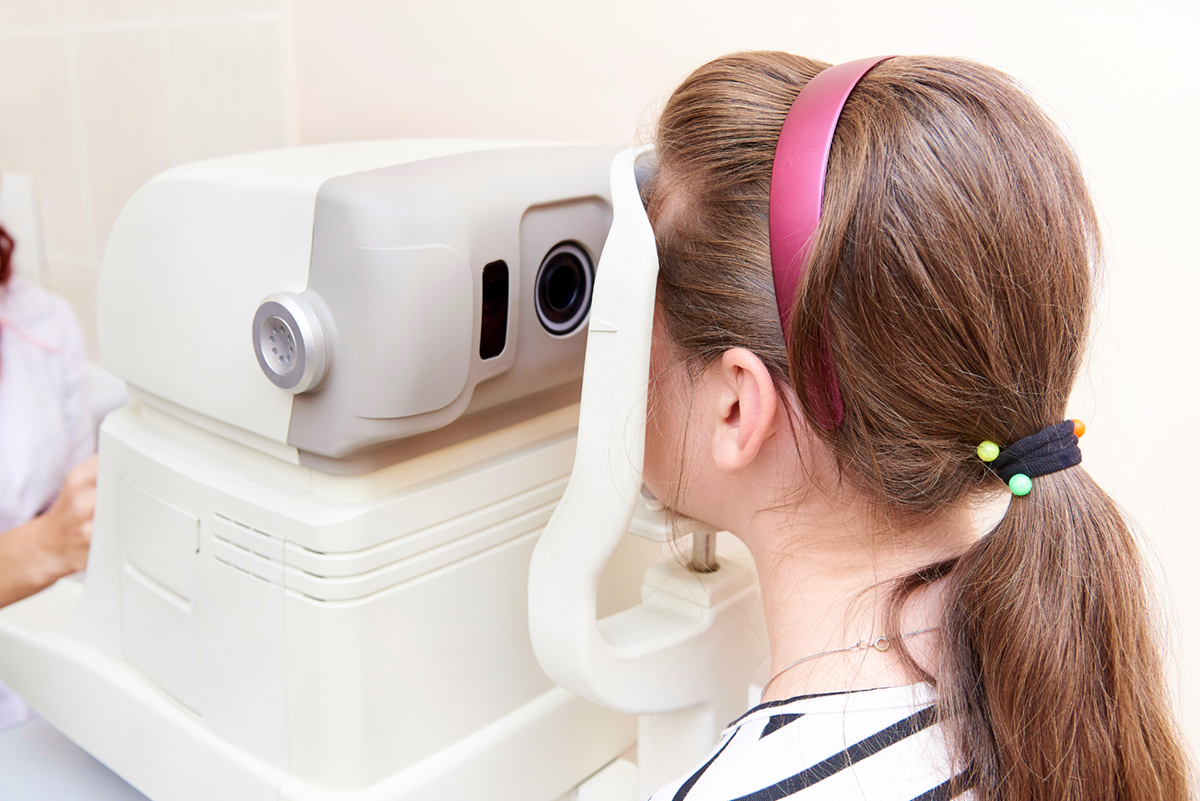
Screening and Diagnosis
Tonometry
Tonometry is a very simple, painless procedure that measures intraocular pressure. Two common techniques are:
- Air-puff tonometry – A puff of air is used to measure the amount of force needed to indent cornea.
- Applanation tonometry – A sophisticated device that's usually fitted to a slit lamp is used.
Test for optic nerve damage
In order to see if the optic nerve has been damaged, the doctor uses ophthalmoscope which enables him or her to look directly through the pupil to the back of your eye.
Perimetry test
This test requires a patient to look at a screen with a target in the center. During this, doctor manipulates a small object on a wand at different locations in your visual field.
Pachymetry
During this test, the doctor uses an ultrasonic wave instrument to gauge the thickness of each cornea. This is because the thickness of corneas is an important factor for accurately diagnosing glaucoma.
If someone has thick corneas, eye pressure may seem high even though he or she doesn't have glaucoma.
Treatment of glaucoma
There are several treatment options for glaucoma. Topical medications are the most common early treatment for glaucoma because standard practice has been to move on to surgery only if medications are ineffective.
Eye drops
There are several types of eye drops that doctors prescribe most commonly.
Beta blockers
These reduce the production of aqueous humor and some of the most commonly used are: levobunolol (Betagan ), timolol (Betimol, Timoptic ), carteolol (Ocupress), betaxolol (Betoptic) and metipranolol (OptiPranolol ).
Alpha-adrenergic agents
These medications also reduce the production of aqueous humor. They include: apraclonidine (Iopidine) and brimonidine (Alphagan).
Carbonic anhydrase inhibitors
These medications, which include dorzolamide (Trusopt), reduce the amount of aqueous humor.
Prostaglandin analogues
These medications are used to increase the outflow of aqueous humor. The most common is latanoprost (Xalatan).
Prostamides
These include bimatoprost (Lumigan). They increase the outflow of aqueous humor.
Miotics
Miotics, such as pilocarpine (Isopto Carpine, Pilocar) increase the outflow of aqueous humor.
Epinephrine compounds
These also increase the outflow of aqueous humor.
Surgery
Trabectomy
During the procedure doctor uses a high-energy laser beam to shrink part of the trabecular meshwork, which causes other parts of the meshwork to stretch and open up. This helps aqueous humor drain more easily from the eye.
Conventional surgery
It is called filtering procedure and it is done in a hospital or an outpatient surgery center. Using delicate instruments under an operating microscope, the doctor creates an opening in the sclera and removes a small piece of the trabecular meshwork. The aqueous humor can now leave the eye freely through this hole.
- www.mayoclinic.com/health/glaucoma/DS00283
- http://www.glaucoma-association.com/nqcontent.cfm?a_id=1476〈=en&tt=article