Table of Contents
Recurrent corneal erosion is a condition affecting the outermost layer of corneal cells called the epithelium. The problem is caused when the bottom layer of epithelial cells adhere poorly to the cornea, causing them to slough off easily.
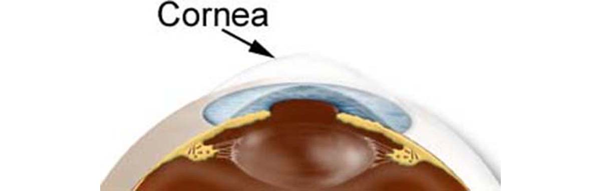
Cornea: Basic anatomy
The cornea is the transparent, dome-shaped window that covers the front of the eye. Although most people don’t know a lot about this part of the eye, it is a powerful refracting surface that provides two thirds of the eye's focusing power. Like the crystal on a watch, the cornea gives us a clear window to look through. The cornea has nerve endings sensitive to touch, temperature and chemicals; a touch of the cornea causes an involuntary reflex to close the eyelid. It is made from several different layers:
-
Corneal epithelium. This is one thin epithelial layer of fast-growing and easily-regenerated cells, kept moist all the time with tears.
- Bowman's layer. This layer is one tough layer that protects the corneal stroma, consisting of irregularly-arranged collagen fibers.
- Corneal stroma. We are talking about one transparent middle layer responsible for most of the focusing that the cornea performs. It is made from regularly-arranged collagen fibers along with fibroblasts. The corneal stroma consists of approximately 200 layers of type I collagen fibrils.
- Posterior limiting membrane. This is a thin acellular layer. The role of this membrane is to be a modified basement membrane of the corneal endothelium
- Corneal endothelium. This is a simple squamous epithelium, which works as a barrier to prevent water inside the eyeball from moving into and hydrating the cornea, which would lead to blurred vision. The corneal endothelium is bathed by aqueous humour, not by blood or lymph, and has a very different origin, function, and appearance from vascular endothelia. The middle of our eye is filled with a jelly-like substance called the vitreous. The vitreous is clear and allows light to pass directly from the front to the back of our eye.
The iris, the colored circle at the front of our eye, changes the size of the pupil which allows different amounts of light into our eye. The pupil is the dark hole in the middle of the colored part of our eye. The pupil gets smaller in bright conditions to let less light in and bigger in dark conditions to let more light in.
Cornea size
In humans, the cornea has a diameter of about 12 mm and a thickness of 0.5 mm - 0.7 mm in the center and 1.0 mm - 1.2 mm at the periphery. Transparency, avascularity, and immunologic privilege makes the cornea a very special tissue. In humans, the refractive power of the cornea is approximately 45 diopters, which amounts to roughly three-fourths of the eye's total power.
Recurrent Corneal Erosion: Incidence of the condition
Recurrent corneal erosions are quite frequently reported in most developing countries, where a lack of proper nutrition plays a significant role in threatening the health of the cornea. If corneal erosions are inherited, the pattern is dominant; however, most corneal erosions are acquired. Recurrent corneal erosions usually are seen as a bilateral (meaning it strikes both eyes) problem occurring somewhat more frequently in females than in males.
This condition occurs in adults, usually after the fourth decade of life. However, there are studies that have associated recurrent corneal erosions with juvenile Alport syndrome and renal complications.
Recurrent Corneal Erosion: Etiology of the condition
Recurrent corneal erosion syndrome is characterized by a disturbance at the level of the corneal epithelial basement membrane, which results in defective adhesions and recurrent breakdowns of the epithelium. Recurrent Corneal Erosion can occur secondary to corneal injury or spontaneously, although some predisposing factors, such as diabetes or a corneal dystrophy may be the underlying cause.
The most common cause of RCE syndrome is definitely trauma to the cornea. In these cases, patients will generally give a history of previous oblique corneal abrasion with an object such as a fingernail, piece of paper… Corneal abrasions may result from:
- foreign bodies
- contact lenses
- chemicals
- fingernails
- hair brushes
- tree branches
- dust
The original injury is generally well recalled by the patient as it is usually followed by several days of pain, watering and photophobia (sensitivity to light). The first recurrence may then not occur for quite a few months after the original trauma. This syndrome can also occur spontaneously, although in this situation there is often some predisposing factor. For example, many corneal dystrophies are associated with recurrent corneal erosions. Other possible causes of Recurrent Corneal Erosion are:
- Post infectious ulcers from herpes simplex
- Exposure
- Cockayne syndrome
- Reis-Bücklers dystrophy
- Vitrectomy
- The improper use of contact lenses
Continue reading after recommendations
- en.wikipedia.org/wiki/Corneal_erosion
- www.emedicine.com
- www.optometry.co.uk