
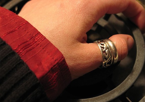
What Is Normal Looking Skin?
- Important notification about information and brand names used in this slideshow!
- Photo courtesy of estrellas spa01 by Picasa : picasaweb.google.com/lh/photo/rSdpRzdeDlQC78ruexh1YA
- Lecture notes on dermatology by Tony Burns and Robin Graham-Brown
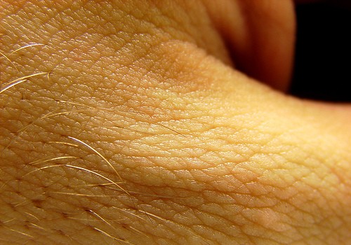
Dry And Cold Skin
- Important notification about information and brand names used in this slideshow!
- Photo courtesy of le-champignon by Flickr : www.flickr.com/photos/le-champignon/133878972/
- Lecture notes on dermatology by Tony Burns and Robin Graham-Brown
Yellow Or Orange Skin
- Important notification about information and brand names used in this slideshow!
- Photo courtesy of Matt Reinbold by Flickr : www.flickr.com/photos/furryscalyman/772243754/
- Lecture notes on dermatology by Tony Burns and Robin Graham-Brown

Rash
- Important notification about information and brand names used in this slideshow!
- Photo courtesy of Arthaey Angosii by Flickr : www.flickr.com/photos/arthaey/2145051891/
- Lecture notes on dermatology by Tony Burns and Robin Graham-Brown
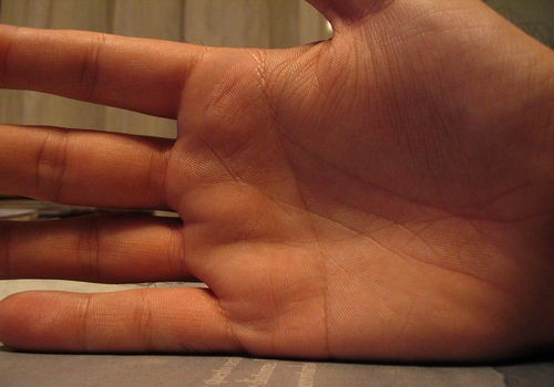
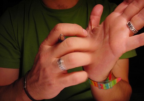
Dark Red Lines On The Palms
- Important notification about information and brand names used in this slideshow!
- Photo courtesy of Sim Dawdler by Flickr : www.flickr.com/photos/simulation/1442156/
- Lecture notes on dermatology by Tony Burns and Robin Graham-Brown
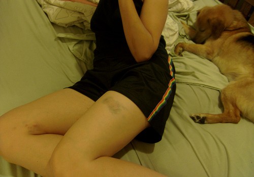
Bruises
- Important notification about information and brand names used in this slideshow!
- Photo courtesy of Thirteen Of Clubs by Flickr : www.flickr.com/photos/thirteenofclubs/2610096635/
- Lecture notes on dermatology by Tony Burns and Robin Graham-Brown
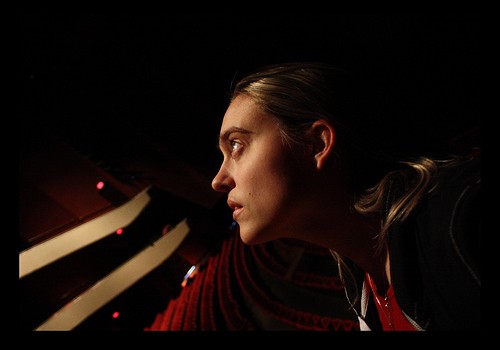
Tingling, Pins And Needles
- Important notification about information and brand names used in this slideshow!
- Photo courtesy of Keturah Stickann by Flickr : www.flickr.com/photos/johnandketurah/3255396640/
- Lecture notes on dermatology by Tony Burns and Robin Graham-Brown

Pale Skin
- Important notification about information and brand names used in this slideshow!
- Photo courtesy of Eddy Van 3000 by Flickr : www.flickr.com/photos/e3000/6234971041/
- Lecture notes on dermatology by Tony Burns and Robin Graham-Brown
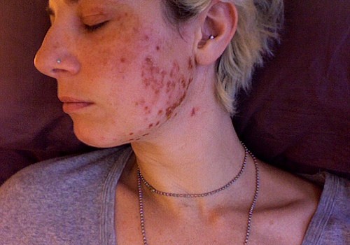
Brown Patches
- Important notification about information and brand names used in this slideshow!
- Photo courtesy of alysha naples by Flickr : www.flickr.com/photos/bluegreen/3703918212/
- Lecture notes on dermatology by Tony Burns and Robin Graham-Brown
Itching Without Cause
- Important notification about information and brand names used in this slideshow!
- Photo courtesy of sergis blog by Flickr : www.flickr.com/photos/srgblog/3951790966/
- Lecture notes on dermatology by Tony Burns and Robin Graham-Brown
Your thoughts on this
Loading...