A "triple A" — an abdominal aortic aneurysm — is a permanent expansion or bulge within the aorta. Although abdominal aortic aneurysms generally don't cause any symptoms, they can become extremely dangerous when left untreated. What do you need to know if you are having an ultrasound screen for an abdominal aortic aneurysm?
How does ultrasound work?
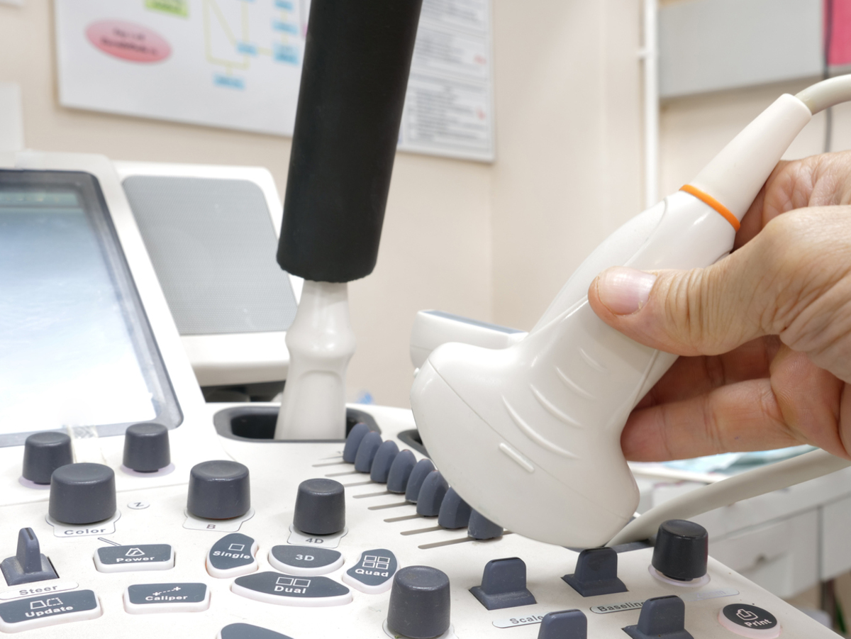
Ultrasound is a painless, non-invasive diagnostic method, which doesn’t use ionizing radiation. It works by using sound waves to produce pictures of structures found in the abdomen. The main part of the ultrasound machine is called a transducer. This transducer, which is held to your skin, emits high frequency ultrasound waves, and then detects a part of those waves that bounce off the internal structures. All of these detected waves are digitally transformed into a real time two-dimensional black-and-white image on the screen.
Unfortunately, water and air do not conduct ultrasound waves that well, so you can’t use ultrasound scans to view the bowels, or to visualize organs covered by the intestines. Other than that, the large amount of tissue in overweight patients tend to weaken the sound waves, making the signal harder to be converted into a 2D picture.
Ultrasound machines can also detect blood flow inside your vessels, using a phenomenon called the “Doppler effect”. Contemporary ultrasound machines can simultaneously show both regular 2D images along with blood flow visualization, using a diagnostic procedure known as “color duplex sonography”.
Do I need to get an ultrasound exam?
Ultrasound helps to diagnose conditions such as:
- Liver and other internal organ issues
- Hernias
- Kidney or gallbladder stones
- Abdominal aortic aneurysm
Symptoms like abdominal pain, circulation problems, or a pulsating mass in the abdominal area are all characteristic of a large abdominal aortic aneurysm. However, due to the lack of symptoms, smaller abdominal aortic aneurysms are usually diagnosed by coincidence while examining other organs in the abdomen.
The main goal of this screening exam is to evaluate the overall condition of the aorta, and to see if there are any potential risks of complications, such as an aortic rupture or dissection.
How do I prepare for an ultrasound scan?
Your doctor will most likely give you a list of things you should and shouldn’t do prior to the examination.
For your convenience, avoid wearing tight clothes when you head to your ultrasound scan, so you can easily uncover your belly area and remove your belly button piercing if you have one. Try to sleep and get some rest the night before the exam. It’s also highly recommended to take a shower before you come. (Although this one should go without saying, I had to write that, because you wouldn’t believe what sights and odors I’ve witnessed during my short career as an intern.)
You may drink water, especially if the doctor would like to see the condition of your bladder. In that case, you should drink four to six glasses of water one hour before the exam.
What is the ultrasound examination like?
An ultrasound exam usually lasts for about 15 to 30 minutes.
First, you need to take off your shirt and take your pants down a bit to expose your belly. Then you lay down on the bed with your face up, and with your hands over your head. Note that your doctor may ask you to turn on your side during the examination, because some abdominal structures are better viewed this way.
Now that you’re in the right position, the doctor will spread water-based gel on the surface of your abdomen. The gel is used to eliminate any potential air space between the transducer and your skin, because ultrasound doesn’t travel well through air. You may feel a slightly cold sensation on your skin at first, but luckily newer transducers come with an option that can warm up the gel.
The transducer is then placed on your body, and the doctor will move it until they acquire the pictures that are needed for the diagnosis. Normally, you can only sense a slight pressure from the transducer, although if the area is sensitive, you might feel some pain.
After the examination is over, and the doctor prints out the images, the gel is wiped off, and you can now dress yourself. Don’t worry if small amounts of gel remain on your skin. Because the gel is water-based, it won’t leave any permanent stains on your clothes.
Finally, you wait outside, while your doctor writes the report. With that report, you can now go to the doctor who asked for the examination, such as a surgeon or a cardiologist, and they will compare these results with their clinical findings, so they can conclude what would be the best therapy for you.
After the examination ends, you can continue with your normal daily activities.
What can the doctor tell me about my aorta?
Every aspect of the abdominal aorta is carefully examined during an utrasound scan, such as:
- The position, length, and the potential variations of the aorta
- The shape and size of the aortic wall
- The blood flow through the aortic lumen
The average size of the abdominal aorta is 1.5 cm in women, and 1.7 cm in men over 50 years old, measured on the level where renal arteries (vessels that supply the kidney) are branching out from the aorta.
The most common location for AAA is just below the renal arteries, and if the aorta is more than 3 cm in diameter, we’re talking about AAA. Aneurysms can grow up to 15 cm in diameter and even up to 25 cm in length. The bigger this diameter, the larger are the chances of a potential aortic rupture.
The shape of the aneurysm is also examined. It can be either fusiform (when equally expanded), and rarely saccular (ball shaped).
When using the duplex sonography method, you can see potential issues in the blood flow due to the narrowing of the aorta, blood clots, or external tumors pressing the blood vessel.