The valves in the heart are responsible for making sure blood flows in the right direction. The pulmonary valve serves the pulmonary artery that sends oxygen-poor blood to the lungs. In pulmonary valve stenosis, a deformity on or near the pulmonary valve causes its opening to narrow, and slowing down blood flow. The valve can either be thickened or fused and therefore, does not fully open.
What causes pulmonary valve stenosis, and what are the risk factors?
Most people who have pulmonary valve stenosis are born with the condition, meaning it is a congenital heart disease. Genetic factors can play a role in its development, and maternal rubella infections during pregnancy also increase the risk a baby will be born with the condition. Its precise cause remains unknown, however.
The currently-known risk factors associated with developing pulmonary valve stenosis later on in life include:
- Carcinoid syndrome
- Rheumatic fever
- Noonan syndrome
- Pulmonary valve replacement
How does pulmonary valve stenosis affect the heart?
Children with pulmonary valve stenosis have elevated pressure in the right side of their heart. Therefore, the heart has to work harder to pump blood into the pulmonary arteries.
What are the symptoms of pulmonary valve stenosis?
The symptoms of pulmonary valve stenosis can vary significantly from person to person depending on the severity of the condition. They can range from non-existent or mild to severe, and are, where present, particularly noticeable during exercise.
Fortunately, mild cases of pulmonary valve stenosis, in which patients don’t have any symptoms, don’t worsen over time. However, patients with moderate or severe pulmonary valve stenosis can deteriorate to the point where they need to undergo surgery.
These are the symptoms associated with pulmonary valve stenosis:
- Heart murmur, an abnormal whooshing sound caused by turbulent blood flow that can be heard through a stethoscope — often leading to diagnosis when a doctor hears it in an infant and sets out to investigate further.
- Fatigue or tiredness.
- Shortness of breath, particularly during exercise.
- Chest pain, known as angina.
- Fainting.
- In severe cases, some babies may have cyanosis, in which they look blue due to a lack of oxygen.
What are the complications associated with pulmonary valve stenosis?
The complications patients with pulmonary valve stenosis are at risk of include:
- Infection. Patients with pulmonary valve stenosis have a higher risk of developing endocarditis, an infection that affects the inner lining of heart.
- A heart that pumps with difficulty. In patients suffering from severe pulmonary valve stenosis, the right side of the heart is forced to work harder to pump blood. This extra work can cause the heart muscle in the right ventricle to thicken, making it stiff and causing it weaken.
- Heart failure. Patients with pulmonary valve stenosis are more likely to develop heart failure, a condition in which the heart doesn’t pump efficiently enough to provide the body with the oxygen it needs.
- Arrhythmia, or an irregular heartbeat.
How is pulmonary valve stenosis diagnosed?
Doctors may suspect pulmonary valve stenosis when they hear a heart murmur through a stethoscope. At that point, they will ask for one of these diagnostic tests to follow up:
- An echocardiogram, a technique that uses sound waves to produce a moving image of a heart.
- An electrocardiogram, which measures the electrical activity of your heart.
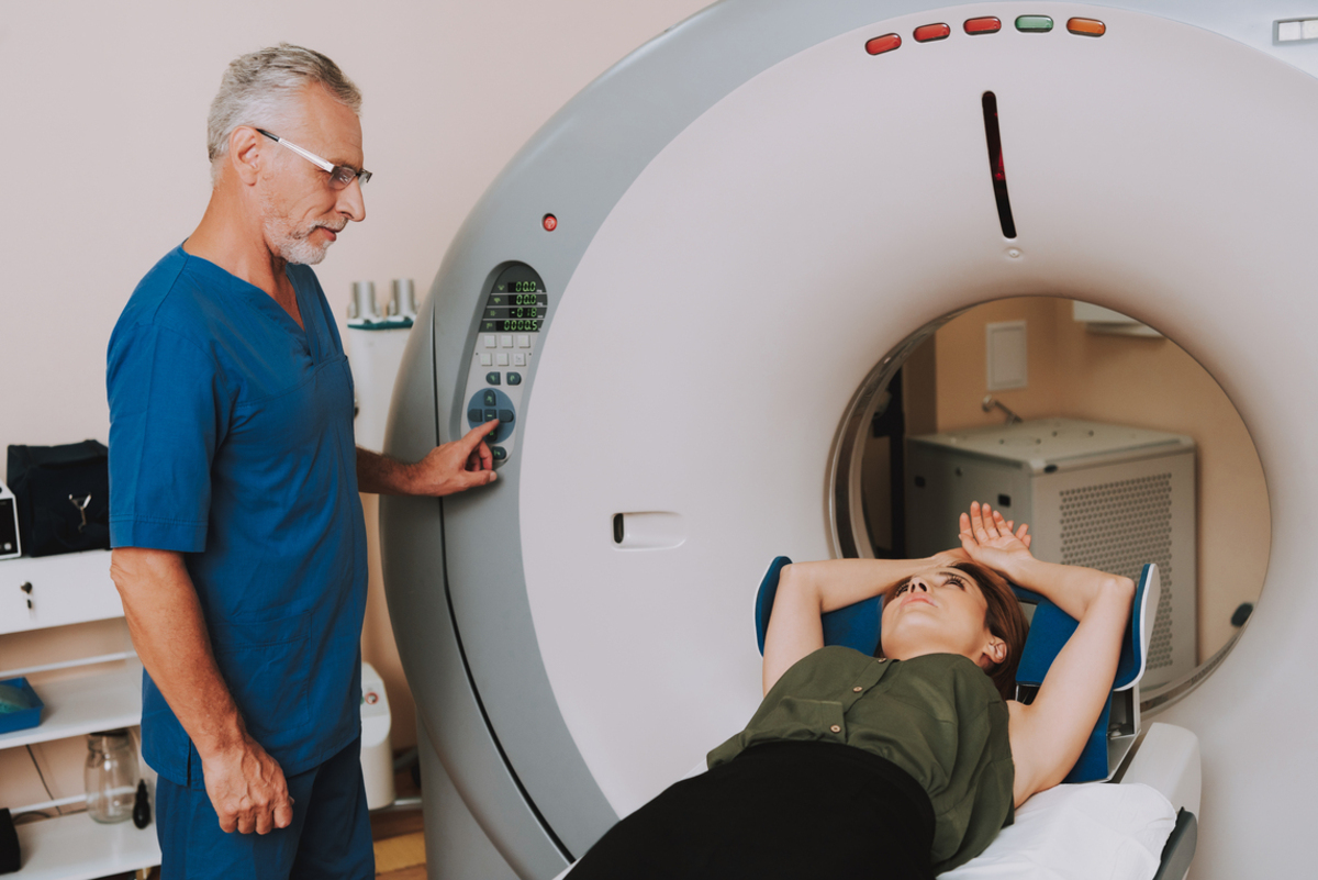
- MRI and CT scans, which are both imaging techniques that help provide detailed views of the heart.
- Cardiac catheterization, which involves inserting a thin and flexible tube up to your heart, thus helping provide a view of your heart and blood vessels.
How is pulmonary valve stenosis treated?
Treatment for pulmonary valve stenosis depends on whether the condition is mild, moderate or severe. Usually, mild cases of the disease don’t cause symptoms and don’t require treatment. However, doctors will keep an eye on the condition through routine checkups. Patients who require treatment will usually have high pressure on the right side of their heart. Their medical teams may recommend:
- A balloon valvuloplasty. This is a technique that is used alongside cardiac catheterization to help relieve the obstruction. In this procedure, a special tool and balloon are placed around the pulmonary valve. The balloon is inflated for some time, which helps to stretch the valve. The balloon is then removed. A common side effect of this procedure is blood leakage.
- Open-heart surgery. Some children with pulmonary valve stenosis will need to undergo surgery. The surgeon will either repair the pulmonary artery or pulmonary valve, or they will replace the valve with an artificial one. Open-heart surgery carries significant risks, including excess bleeding, infection, and blood clots, but can improve the prognosis.
Treatments for complications of pulmonary valve stenosis
Some treatments can help prevent complications, such as:
- Preventive antibiotics, which can help prevent the development of endocarditis.
- Living a heart-healthy lifestyle, such as quitting smoking, eating a heart-healthy diet, maintaining a healthy weight and doing regular physical activity.
- Kan, J. S., White Jr, R. I., Mitchell, S. E., & Gardner, T. J. (1982). Percutaneous balloon valvuloplasty: a new method for treating congenital pulmonary-valve stenosis. New England Journal of Medicine, 307(9), 540-542.
- Sullivan, I. D., Robinson, P. J., Macartney, F. J., Taylor, J. F., Rees, P. G., Bull, C., & Deanfield, J. E. (1985). Percutaneous balloon valvuloplasty for pulmonary valve stenosis in infants and children. Heart, 54(4), 435-441.
- Walls, J. T., Lababidi, Z., Curtis, J. J., & Silver, D. (1984). Assessment of percutaneous balloon pulmonary and aortic valvuloplasty. The Journal of Thoracic and Cardiovascular Surgery, 88(3), 352-356.
- Photo courtesy of SteadyHealth
Your thoughts on this