Individuals with snapping hip syndrome may feel it when they walk, get up from a chair or swing their leg around. There are several causes for snapping hip syndrome, most commonly due to tendons catching on bony prominences. It is also known as “coxa saltans” syndrome.
Incidence of the condition
Unfortunately, no data are available on the prevalence or incidence of snapping hip syndrome. The fact is that the syndrome occurs most often in individuals aged 15-40 years and affects females slightly more often than males. In one clinical study, the rate of some form of snapping hip syndrome in female ballet dancers with hip complaints was 43.8%, and approximately 30% noted pain with this condition. Athletes, such as ballet dancers and gymnasts, are at special risk for snapping hip syndrome due to repetitive and physically demanding movements.
 Anatomy of the hip joint
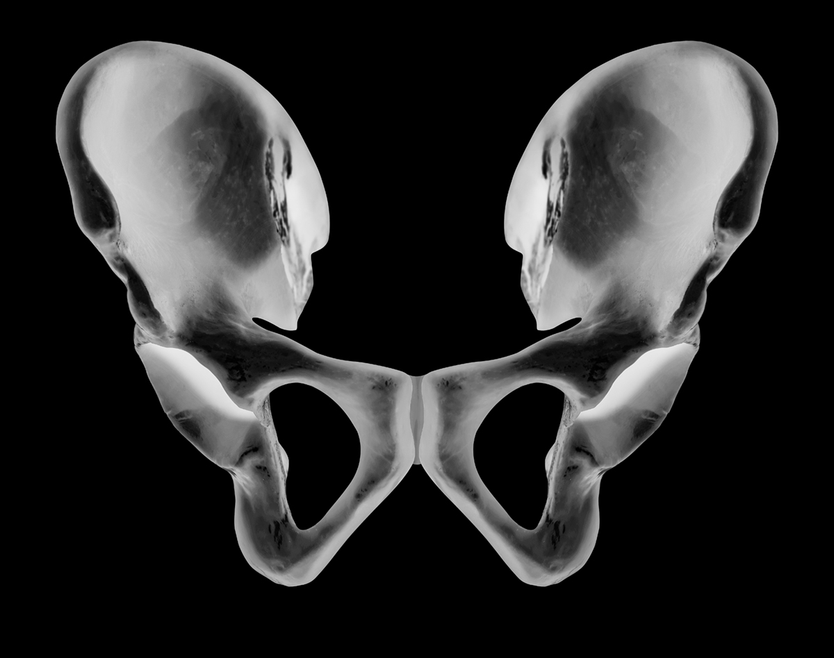
What exactly is the pelvis from the anatomy perspective? It is the link between the trunk and the lower extremities. There is a hip joint between these entities.
Normal movements in this joint include:
- 3° of freedom,
- approximately 120° of flexion,
- 20° of extension,
- 40° of abduction,
- 25° of adduction, and
- 45° each of internal and external rotation.
What strengthens this joint?
The ligament called the ilio-tibial band, or tensor fascia lata. It is a ligament that originates from the iliac crest and inserts on the lateral proximal tibia. Crossing 2 joints, this ligament functions to flex and rotate the thigh medially.
Possible causes of snapping hip syndrome
Several studies have proven that there are three primary causes for snapping hip syndrome:
- Internal - Due to tight hip flexor muscle/tendon called the Iliopsoas muscle/tendon)
- External - Due to tight Iliotibial band that passes over the outside of the hip over the large bump called the greater trochanter of the femur bone.
- Intra-articular (uncommon).A tear in the cartilage or some bone debris in the hip joint can also causes a snapping or clicking sensation. This type of snapping hip usually causes pain and may be disabling. A loose piece of cartilage can cause the hip to catch or lock up
Iliotibial Band Snap
What exactly is the Iliotibial band? The iliotibial band is a thick, wide tendon over the outside of the hip joint. Most people do not know that it represents the most common cause of snapping hip syndrome.? Iliotibial band snaps over the greater trochanter - bone prominence over the outside of the hip joint. Important fact is that, if this is the cause of snapping hip syndrome, patients may develop trochanteric bursitis from the irritation of the bursa-part of the joint, in this region.
Iliopsoas Tendon Snap
The iliopsoas tendon is the primary hip flexor muscle, and the tendon of this muscle passes just in front of the hip joint. The problem is that this tendon can catch on a bony prominence of the pelvis and cause a snap when the hip is flexed. Usually when the iliopsoas tendon is the cause of snapping hip syndrome, patients have no problems, but may find the snapping annoying and hard to handle.
Hip Labral Cartilage Tear
The least common cause of snapping hip syndrome is a tear of the cartilage in the hip joint. Several researches have confirmed that if there is a loose flap of cartilage catching within the joint, this may cause a snapping sensation when the hip is moved. This cause of snapping hip syndrome may also cause an unsteady feeling, and patients may grab for support when the hip snaps.
Symptoms of snapping hip syndrome
There are several possible symptoms that could indicate snapping hip syndrome.
Audible snap
Most commonly, individuals report an audible snap or click in the hip, which may be either painless or painful. The location may be described as lateral or anterior and deep in the groin.
Hip popping out sensations
Occasionally, the phenomenon of a sensation of the hip subluxing or going out of place is felf and is associated with the iliotibial band.
Groin pain
Patients reporting anterior groin pain usually note that the pain is dull or aching in nature and is exacerbated by extension of the flexed, abducted, and externally rotated hip. The pain and snapping may subside with decreased activity and rest. The duration of symptoms at presentation is several months or years rather than days or weeks.
Diagnosis of snapping hip syndrome
Inspection
Every doctor should first examine the gait for abnormalities in biomechanics. Normally if associated iliopsoas tendonitis (inflammation of the tendons) is present, the patient may have a flexed knee. Then joint should be observed as well as the production of the snapping. Some symptoms could be mild and trivial, but external snapping hip syndrome associated with subluxation of the iliotibial band over the greater trochanter may be dramatic and appear as if the patient is subluxing his or her hip.
Palpation
Palpation should definitely be the next step in diagnosis of this condition. Those with external snapping hip syndrome may have tenderness over the proximal iliotibial band, lateral margin of the gluteus maximus. Patients with internal type of the condition may demonstrate an anterior pelvic tilt. Snapping occurs with extension of the flexed, abducted, and externally rotated hip.
Functional testing
There are several functional testings available for the proper diagnosis. External type of the condition is often reproduced with passive internal and external rotation of the hip with the patient in the side-lying position. Internal snapping hip syndrome symptoms can be reproduced with extension of the flexed (30°), abducted, and externally rotated hip.
Imaging diagnostics: X-ray, MRI, ultrasound…
An X-Ray is usually taken to confirm that there is no bony problem around the hip joint, but there is one problem - X-Rays are almost always normal with snapping hip syndrome. If the cause of snapping hip syndrome is thought to be due to a tear of the cartilage within the hip joint, an MRI may be obtained to look for evidence of this problem which is difficult to diagnose.
Ultrasound is a useful, noninvasive diagnostic adjunct because it may demonstrate changes in anatomy and provide an assessment of function. The great benefit of the ultrasound in the case of snapping hip syndrome is the ability to visualize the iliopsoas tendon to provide a directed injection of anesthetic and possible subsequent pain relief.
Lab Studies
There are no specific laboratory studies. For an individual in whom the hip pain is of an unclear origin or with imaging study results suggestive of other pathology, specific consideration should be given for diagnostic laboratory studies at that time.
Treatments of snapping hip syndrome
Physical therapy
Treatment for each patient with snapping hip syndrome should begin with the appropriate physical therapy! Biomechanical assessment of the patient includes both static (posture) and dynamic elements. Particular areas of attention during this portion of the examination include
- knee flexion contracture,
- overpronation of the foot,
- hip flexion contracture, a
- internal or external rotation
Treatment during the acute phase consists of standard anti-inflammatory care and the elimination of activities that exacerbate symptoms.
Ultrasound, phonophoresis, electrical stimulation, iontophoresis can also be used.
Medications
Occasionally, patients may require intermittent Non-steroidal anti Inflammation drugs therapy or simple analgesics as they progress in activities. A corticosteroid injection may be beneficial for those who have persistent pain despite an adequate therapy program.
Surgery
There are several surgical interventions that have been described for patients with persistent pain associated with a snapping hip syndrome. Of course, they should be considered only if the conservative treatment has failed. An operative approach involving a partial release and lengthening of the iliopsoas tendon, with minimal resection of a lesser trochanteric bony ridge, if involved, is described. However, every patient should note that surgical intervention is rarely necessary in the management of this condition.
- Resection of the posterior half of the iliotibial tract at the insertion site of the gluteus maximus, with excision of the trochanteric bursa.
- Elliptical resection of a portion of the iliotibial band overlying the greater trochanter, with removal of the trochanteric bursa, can be performed.
- Z-plasty of the iliotibial band, resulting in lengthening of the tendon, is another option.
- A lengthening procedure can be performed on the iliopsoas tendon, typically by partial release of the tendon
- Resection of the bony prominence of the lesser trochanter.
- Complete release of the iliopsoas tendon.
Prognosis
This condition is usually curable with appropriate treatment, or sometimes it heals spontaneously. If it is painless, there is little cause for concern. If it is bothersome or painful, stretching of the tight structures can alleviate the symptoms in 6-8 weeks of consistent attention.
- www.orthopedics.about.com
- www.sportsinjurybulletin.com
- www.emedicine.com
- en.wikipedia.org/wiki/Snapping_hip_syndrome