Table of Contents
The Diagnosis of Hip Dysplasia
A family history of hip dysplasia or extremely flexible ligaments, being a breech baby, and orthopedic problems including club foot, are risk factors for hip dysplasia. Girls are affected more often than boys, and firstborns are at a higher risk as well. Hip dysplasia is routinely screened for during infancy in some countries, while other countries prefer to screen only if certain risk factors are present.
Initially, a baby's hips may be moved around by a pediatrician to check if a questionable “click” can be heard. If so, further screening will involve ultrasound or X-ray pictures — often at three months of age. These diagnostic measures will determine the extent of hip dysplasia as well as enabling a diagnosis.
This is why the International Hip Dysplasia Institute describes hip dysplasia as a “silent condition”. But the fact that hip dysplasia is highly unlikely to form a problem for your infant does not mean that the condition is harmless. Hip pain, a limp, and hip joints that wear out at a much faster rate than normal can be the result in adults.
Early detection and treatment can usually prevent these problems, so facilitating those steps is important. If you live in a country that doesn't pay meticulous attention to their infants' hips (like many European countries do), there is nothing to stop you from asking your pediatrician to check your baby's hips during a routine well-baby visit, especially if your baby has certain risk factors for hip dysplasia.
Parents, too, can play an important role in the diagnostic process. Symptoms of hip dysplasia you may notice in your infant include:
-
Asymmetrical creases in the skin of your baby's buttocks.
-
One leg appears to be shorter than the other.
-
The hip or hips click or pop — though this can be due to other conditions as well.
-
The baby's legs may not be able to spread fully, which you may notice during diaper changes.
-
A limp, or “waddling like a duck” may be noticed after the baby starts walking.
Treatment Options for Hip Dysplasia
Non-surgical treatment options that aim to push the hip into a better position and try to enable correct hip joint development are most common in infants under the age of six months. There are several methods available, and the chosen method often depends on the country in which treatment is conducted.
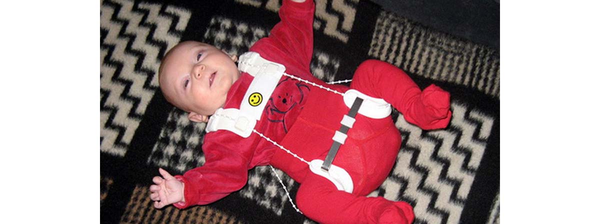
A Pavlik Harness vaguely resembles weird dungarees. It keeps the hips and knees bent and the legs spread wide apart. Other, less flexible harnesses can also be used. A Frejka pillow is a big pillow held up by straps that serves the same purpose. Traction is also used to stretch the ligaments, particularly in Asia and Europe, and usually before surgery. This involves holding the legs up with a device, or placing a pillow under the legs and applying traction length-ways. Traction is controversial, because it is not clear if it actually helps to promote proper hip development in infants.
A closed reduction is usually performed on children aged 24 months or younger. This operation is done under general anesthesia. After cutting the abductor tendon, the surgeon pushes the “ball” of the upper thigh bone back into normal position. The child is then placed into a so-called spica cast to force the hip to stay in proper alignment.
In an open reduction, tissues that prevent the ball (femur) from going into the socket are removed. There are two variations of this procedure, suitable for different ages and situations. Sometimes, it involves reshaping the hip socket.
Osteotomy refers to the reshaping of a bone. A pelvic osteotomy involves the reshaping of the hip socket, while a femoral osteotomy refers to reshaping the femur, or ball of the thigh bone, which needs to fit into the hip socket. Both these procedures may also be combined with either open or closed reductions.
- Photo courtesy of Pedro Simões by Wikimedia Commons : commons.wikimedia.org/wiki/File:Hip_horizon.jpg
- Photo courtesy of Thiemo Schuff by Wikimedia Commons : commons.wikimedia.org/wiki/File:Saeugling_mit_angelegter_spreizhose.jpg
- Photo courtesy of Hip2PAOFO by Wikimedia Commons : commons.wikimedia.org/wiki/File:FO_after.jpg
- en.wikipedia.org/wiki/Hip_dysplasia_(human)
- http://www.hipdysplasia.org/
- http://pediatrics.aappublications.org/content/105/4/896
- www.mayoclinic.org/hip-dysplasia/
Your thoughts on this