The L6 vertebra
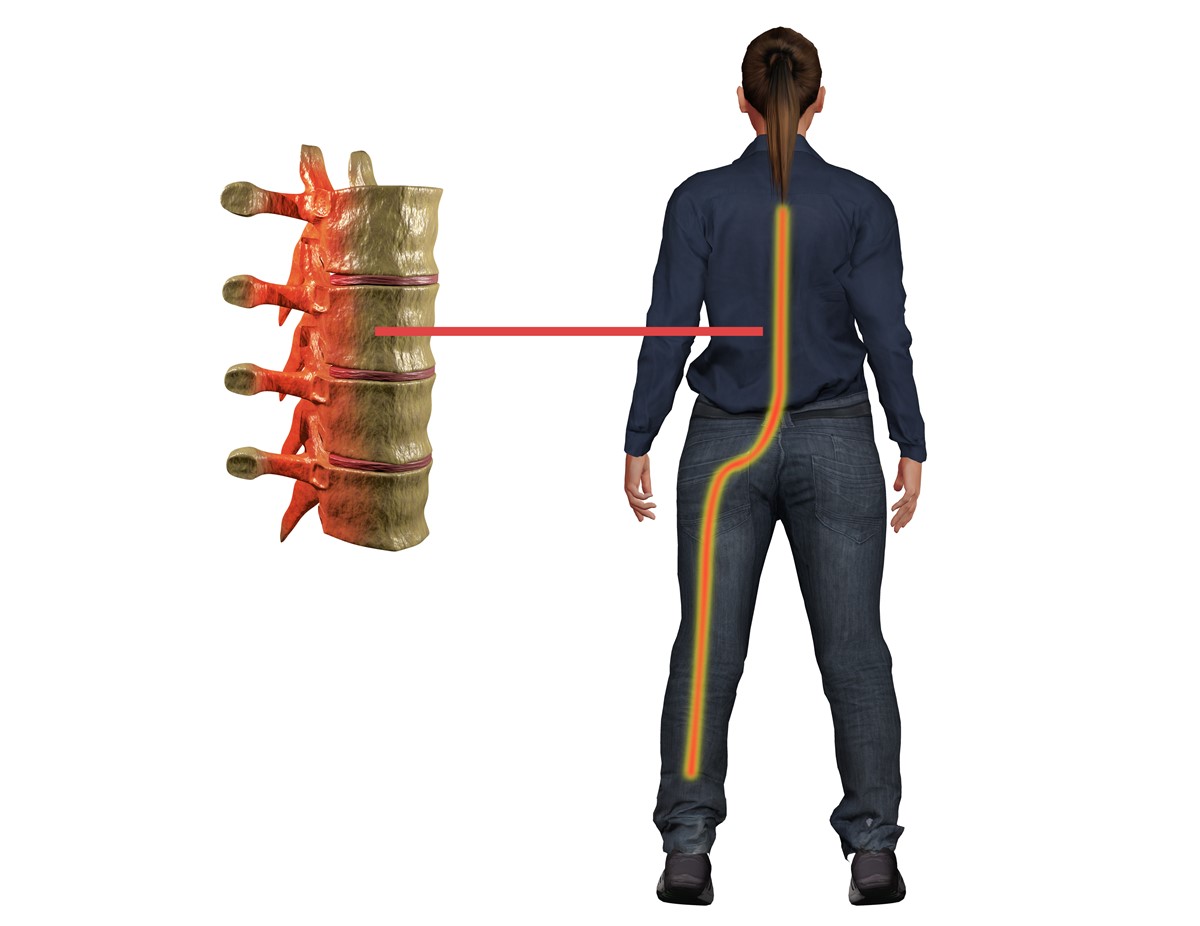
The lumbar region of the spine, also known as the lower back, consists of 5 bones or vertebrae. These bones are given a numerical sequence and named L1 to L5. In 10% of adults a spinal abnormality can occur due to genetics and this can cause the presence of an extra or transitional vertebra called L6.
The presence of this extra vertebrae doesn't necessarily cause any more problematic spinal conditions that would occur in those who don't have the extra bone. The L6 vertebra is subject to the exact same pathology as patients would experience at the L5 vertebra. Any pathology at the L6 vertebrae is referred to as an issue at the L6-S1 level rather than L5-L6 levels. This is to emphasize that L6 is associated with the sacral region which is situated below the lumbar region of the back.
Specific L6 vertebra conditions
Other conditions which can affect this vertebra include a herniated disk, spinal stenosis, a bulging disk, osteoarthritis and degenerative disk disease. In any case, these are all conditions which can affect any level of the spine.
Investigations and management
In most cases, the diagnosis of an L6 vertebra is coincidental. Patients may complain of lower back pain due to mechanical issues or an acute injury and X-rays of the lower back will then show the presence of the extra vertebra.
Back hygiene is very important and these tips should be followed to help alleviate lower back pain.
- Walking and swimming are good activities to help with strengthening of the back muscles which then offer support to the lower spine.
- When performing activities or lifting up heavy weights, always bend the knees and hips and not the back.
- Try losing some extra kilograms if overweight as this will relieve pressure on the back.
- Maintain a good posture by keeping a straight back when sitting and standing.
If the patient is experiencing any neurological signs, such as weakness or decreased sensation in the body or the limbs, or the patient's pain is getting worse despite conservative management, then a CT scan and/or MRI scan will be done to investigate and rule out any other possible pathology.
In the event that conservative management fails completely or there is pathology, such as those mentioned above, then surgical intervention may be considered.
Conclusion
- Photo courtesy of SteadyHealth